 Movie
Movie Controller


+ Open data

- Basic information
| Entry | Database: PDB / ID: 1dgl | |||||||||
|---|---|---|---|---|---|---|---|---|---|---|
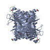
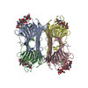
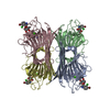
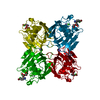
| Title | LECTIN FROM DIOCLEA GRANDIFLORA COMPLEXED TO TRIMANNOSIDE | |||||||||
 Components Components | LECTIN | |||||||||
 Keywords Keywords | LECTIN / SACCHARIDE BINDING / TRIMANNOSIDE | |||||||||
| Function / homology |  Function and homology information Function and homology informationD-mannose binding / toxin activity / carbohydrate binding / metal ion binding / identical protein binding Similarity search - Function | |||||||||
| Biological species |  Dioclea grandiflora (mucana) Dioclea grandiflora (mucana) | |||||||||
| Method |  X-RAY DIFFRACTION / MOLECULAR REPLACEMENT / Resolution: 2.4 Å X-RAY DIFFRACTION / MOLECULAR REPLACEMENT / Resolution: 2.4 Å | |||||||||
 Authors Authors | Rozwarski, D.A. / Swami, B.M. / Brewer, C.F. / Sacchettini, J.C. | |||||||||
 Citation Citation | Journal: J.Biol.Chem. / Year: 1998 Title: Crystal structure of the lectin from Dioclea grandiflora complexed with core trimannoside of asparagine-linked carbohydrates. Authors: Rozwarski, D.A. / Swami, B.M. / Brewer, C.F. / Sacchettini, J.C. #1: Journal: J.Biol.Chem. / Year: 1996Title: Structural Basis of Trimannoside Recognition by Concanavalin A Authors: Naismith, J.H. / Field, R.A. | |||||||||
| History |
|
- Structure visualization
| Structure viewer | Molecule: MolmilJmol/JSmol |
|---|
- Downloads & links
-Download
| PDBx/mmCIF format | 1dgl.cif.gz | 108.9 KB | Display | PDBx/mmCIF format |
|---|---|---|---|---|
| PDB format | pdb1dgl.ent.gz | 84.2 KB | Display | PDB format |
| PDBx/mmJSON format | 1dgl.json.gz | Tree view | PDBx/mmJSON format | |
| Others |  Other downloads Other downloads |
-Validation report
| Arichive directory | https://data.pdbj.org/pub/pdb/validation_reports/dg/1dglftp://data.pdbj.org/pub/pdb/validation_reports/dg/1dgl | HTTPS FTP |
|---|
-Related structure data
| Related structure data |  5cnaS S: Starting model for refinement |
|---|---|
| Similar structure data |


-Links
 PDBj
PDBj

- Assembly
| Deposited unit | 
| ||||||||
|---|---|---|---|---|---|---|---|---|---|
| 1 | 
| ||||||||
| Unit cell |
| ||||||||
| Noncrystallographic symmetry (NCS) | NCS oper: (Code: given Matrix: (0.496, 0.864, -0.087), Vector: |
-Components
| #1: Protein | Mass: 25572.285 Da / Num. of mol.: 2 / Source method: isolated from a natural source / Details: TRIMANNOSIDE COMPLEX / Source: (natural) Dioclea grandiflora (mucana) / References: UniProt: P08902#2: Polysaccharide | Source method: isolated from a genetically manipulated source #3: Chemical |   Mass: 40.078 Da / Num. of mol.: 2 / Source method: obtained synthetically / Formula: Ca Mass: 40.078 Da / Num. of mol.: 2 / Source method: obtained synthetically / Formula: Ca#4: Chemical |   Mass: 54.938 Da / Num. of mol.: 2 / Source method: obtained synthetically / Formula: Mn Mass: 54.938 Da / Num. of mol.: 2 / Source method: obtained synthetically / Formula: Mn#5: Water | ChemComp-HOH / |  Mass: 18.015 Da / Num. of mol.: 190 / Source method: isolated from a natural source / Formula: H2O Mass: 18.015 Da / Num. of mol.: 190 / Source method: isolated from a natural source / Formula: H2O |
|---|
-Experimental details
-Experiment
| Experiment | Method: X-RAY DIFFRACTION / Number of used crystals: 1 |
|---|
- Sample preparation
| Crystal | Density Matthews: 4.85 Å3/Da / Density % sol: 75 % | ||||||||||||||||||||||||||||||||||||
|---|---|---|---|---|---|---|---|---|---|---|---|---|---|---|---|---|---|---|---|---|---|---|---|---|---|---|---|---|---|---|---|---|---|---|---|---|---|
| Crystal grow | pH: 7.2 / Details: pH 7.2 | ||||||||||||||||||||||||||||||||||||
| Crystal grow | *PLUS Temperature: 4 ℃ / Method: vapor diffusion, hanging drop | ||||||||||||||||||||||||||||||||||||
| Components of the solutions | *PLUS
|
-Data collection
| Diffraction | Mean temperature: 298 K |
|---|---|
| Diffraction source | Source: ROTATING ANODE / Type: RIGAKU RUH2R / Wavelength: 1.5418 |
| Detector | Type: SIEMENS / Detector: AREA DETECTOR / Date: Jun 1, 1995 |
| Radiation | Monochromatic (M) / Laue (L): M / Scattering type: x-ray |
| Radiation wavelength | Wavelength: 1.5418 Å / Relative weight: 1 |
| Reflection | Resolution: 2.4→10 Å / Num. obs: 29915 / % possible obs: 74 % / Redundancy: 3.7 % / Rsym value: 0.067 / Net I/σ(I): 13 |
| Reflection shell | Resolution: 2.4→2.5 Å / Redundancy: 1.5 % / Mean I/σ(I) obs: 2.6 / Rsym value: 0.21 / % possible all: 23 |
| Reflection | *PLUS Highest resolution: 2.6 Å / Num. obs: 26679 / Redundancy: 3.9 % / Rmerge(I) obs: 0.064 |
| Reflection shell | *PLUS Highest resolution: 2.6 Å / Lowest resolution: 2.8 Å / % possible obs: 70.5 % / Redundancy: 3.4 % / Num. unique obs: 4349 / Rmerge(I) obs: 0.214 / Mean I/σ(I) obs: 3.8 |
- Processing
| Software |
| ||||||||||||||||||||||||||||||||||||||||||||||||||||||||||||||||||||||||||||||||
|---|---|---|---|---|---|---|---|---|---|---|---|---|---|---|---|---|---|---|---|---|---|---|---|---|---|---|---|---|---|---|---|---|---|---|---|---|---|---|---|---|---|---|---|---|---|---|---|---|---|---|---|---|---|---|---|---|---|---|---|---|---|---|---|---|---|---|---|---|---|---|---|---|---|---|---|---|---|---|---|---|---|
| Refinement | Method to determine structure: MOLECULAR REPLACEMENT Starting model: 5CNA Resolution: 2.4→10 Å / Data cutoff high absF: 100000 / Data cutoff low absF: 0.1 / Isotropic thermal model: RESTRAINED / Cross valid method: THROUGHOUT / σ(F): 2
| ||||||||||||||||||||||||||||||||||||||||||||||||||||||||||||||||||||||||||||||||
| Refinement step | Cycle: LAST / Resolution: 2.4→10 Å
| ||||||||||||||||||||||||||||||||||||||||||||||||||||||||||||||||||||||||||||||||
| Refine LS restraints |
| ||||||||||||||||||||||||||||||||||||||||||||||||||||||||||||||||||||||||||||||||
| LS refinement shell | Resolution: 2.4→2.5 Å / Total num. of bins used: 8
| ||||||||||||||||||||||||||||||||||||||||||||||||||||||||||||||||||||||||||||||||
| Xplor file |
| ||||||||||||||||||||||||||||||||||||||||||||||||||||||||||||||||||||||||||||||||
| Software | *PLUS Name: X-PLOR / Version: 3.1 / Classification: refinement | ||||||||||||||||||||||||||||||||||||||||||||||||||||||||||||||||||||||||||||||||
| Refinement | *PLUS Num. reflection obs: 26679 / Rfactor obs: 0.182 / Rfactor Rfree: 0.247 / Highest resolution: 2.6 Å | ||||||||||||||||||||||||||||||||||||||||||||||||||||||||||||||||||||||||||||||||
| Solvent computation | *PLUS | ||||||||||||||||||||||||||||||||||||||||||||||||||||||||||||||||||||||||||||||||
| Displacement parameters | *PLUS | ||||||||||||||||||||||||||||||||||||||||||||||||||||||||||||||||||||||||||||||||
| Refine LS restraints | *PLUS
| ||||||||||||||||||||||||||||||||||||||||||||||||||||||||||||||||||||||||||||||||
| LS refinement shell | *PLUS Rfactor Rfree: 0.338 / Num. reflection obs: 4349 / Rfactor obs: 0.28 |
