 Movie
Movie Controller

[English] 日本語
 Yorodumi
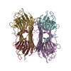
- PDB-1cn1: CRYSTAL STRUCTURE OF DEMETALLIZED CONCANAVALIN A. THE METAL-BINDI... -
+ Open data

- Basic information
| Entry | Database: PDB / ID: 1cn1 | ||||||
|---|---|---|---|---|---|---|---|
| Title | CRYSTAL STRUCTURE OF DEMETALLIZED CONCANAVALIN A. THE METAL-BINDING REGION | ||||||
 Components Components | CONCANAVALIN A | ||||||
 Keywords Keywords | LECTIN (AGGLUTININ) | ||||||
| Function / homology |  Function and homology information Function and homology informationregulation of defense response to virus / D-mannose binding / defense response / metal ion binding Similarity search - Function | ||||||
| Biological species |   Canavalia ensiformis (jack bean) Canavalia ensiformis (jack bean) | ||||||
| Method |  X-RAY DIFFRACTION / Resolution: 3.2 Å X-RAY DIFFRACTION / Resolution: 3.2 Å | ||||||
 Authors Authors | Shoham, M. / Yonath, A. / Sussman, J.L. / Moult, J. / Traub, W. / Gilboa(Kalb), A.J. | ||||||
 Citation Citation | Journal: J.Mol.Biol. / Year: 1979 Title: Crystal structure of demetallized concanavalin A: the metal-binding region. Authors: Shoham, M. / Yonath, A. / Sussman, J.L. / Moult, J. / Traub, W. / Kalb, A.J. | ||||||
| History |
|
- Structure visualization
| Structure viewer | Molecule: MolmilJmol/JSmol |
|---|
- Downloads & links
-Download
| PDBx/mmCIF format | 1cn1.cif.gz | 77 KB | Display | PDBx/mmCIF format |
|---|---|---|---|---|
| PDB format | pdb1cn1.ent.gz | 52.6 KB | Display | PDB format |
| PDBx/mmJSON format | 1cn1.json.gz | Tree view | PDBx/mmJSON format | |
| Others |  Other downloads Other downloads |
-Validation report
| Arichive directory | https://data.pdbj.org/pub/pdb/validation_reports/cn/1cn1ftp://data.pdbj.org/pub/pdb/validation_reports/cn/1cn1 | HTTPS FTP |
|---|
-Related structure data
| Similar structure data |
|---|


-Links
 PDBj
PDBj

- Assembly
| Deposited unit | 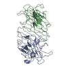
| ||||||||
|---|---|---|---|---|---|---|---|---|---|
| 1 | 
| ||||||||
| Unit cell |
|
-Components
| #1: Protein | Mass: 25596.299 Da / Num. of mol.: 2 Source method: isolated from a genetically manipulated source Source: (gene. exp.) Canavalia ensiformis (jack bean) / References: UniProt: P02866 |
|---|
-Experimental details
-Experiment
| Experiment | Method: X-RAY DIFFRACTION |
|---|
- Sample preparation
| Crystal | Density Matthews: 2.29 Å3/Da / Density % sol: 46.36 % |
|---|---|
| Crystal grow | *PLUS Method: otherDetails: Jack, A., (1971) J. Mol. Biol., 58, 389., Kalb, A.J., (1968) Biochem. J., 109, 669. |
-Data collection
| Radiation | Scattering type: x-ray |
|---|---|
| Radiation wavelength | Relative weight: 1 |
- Processing
| Refinement | Highest resolution: 3.2 Å Details: TWO GROUP TEMPERATURE FACTORS ARE ASSIGNED TO RESIDUES, ONE FOR BACKBONE ATOMS AND THE OTHER FOR SIDE CHAIN ATOMS. | ||||||||||||
|---|---|---|---|---|---|---|---|---|---|---|---|---|---|
| Refinement step | Cycle: LAST / Highest resolution: 3.2 Å
| ||||||||||||
| Refinement | *PLUS Lowest resolution: 10 Å / Num. reflection obs: 7801 / Rfactor obs: 0.256 | ||||||||||||
| Solvent computation | *PLUS | ||||||||||||
| Displacement parameters | *PLUS |
