 Movie
Movie Controller
 Structure viewers
 About EMN search


-Search query
-Search result
Showing 1 - 50 of 142 items for (author: zyla & d)

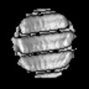
EMDB-72146: 
Structure of the Measles virus Hemagglutinin ectodomain in complex with neutralizing antibody 1C02
Method: single particle / : Zyla D, Acciani M, Niemeyer G, Saphire EO

EMDB-72147:
Structure of the Measles virus Hemagglutinin ectodomain in complex with neutralizing antibody 1C08
Method: single particle / : Zyla D, Acciani M, Niemeyer G, Saphire EO

EMDB-72149:
Structure of the Measles virus Hemagglutinin ectodomain in complex with two neutralizing antibodies 1C08 and 4D08
Method: single particle / : Zyla D, Acciani M, Niemeyer G, Saphire EO

EMDB-72151:
Structure of the Measles virus Fusion glycoprotein ectodomain in complex with two neutralizing antibodies 4F09 and 3A12
Method: single particle / : Zyla D, Acciani M, Niemeyer G, Saphire EO

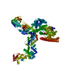
PDB-9q1z:
Structure of the Measles virus Hemagglutinin ectodomain in complex with neutralizing antibody 1C02
Method: single particle / : Zyla D, Acciani M, Niemeyer G, Saphire EO

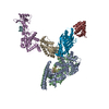
PDB-9q20:
Structure of the Measles virus Hemagglutinin ectodomain in complex with neutralizing antibody 1C08
Method: single particle / : Zyla D, Acciani M, Niemeyer G, Saphire EO

PDB-9q24:
Structure of the Measles virus Hemagglutinin ectodomain in complex with two neutralizing antibodies 1C08 and 4D08
Method: single particle / : Zyla D, Acciani M, Niemeyer G, Saphire EO

PDB-9q26:
Structure of the Measles virus Fusion glycoprotein ectodomain in complex with two neutralizing antibodies 4F09 and 3A12
Method: single particle / : Zyla D, Acciani M, Niemeyer G, Saphire EO

EMDB-72198:
Negative Stain Electron Microscopy map of the Measles Virus Hemagglutinin Glycoprotein Ectodomain in Complex with the Neutralizing Antibody 1G03
Method: single particle / : Zyla D, Acciani M, Niemeyer G, Saphire EO

EMDB-72179:
Negative Stain Electron Microscopy map of the Measles Virus Fusion Glycoprotein Ectodomain in Complex with the Neutralizing Antibody 2B11
Method: single particle / : Zyla D, Acciani M, Niemeyer G, Saphire EO

EMDB-72181:
Negative Stain Electron Microscopy map of the Measles Virus Fusion Glycoprotein Ectodomain in Complex with the Neutralizing Antibody 2G05
Method: single particle / : Zyla D, Acciani M, Niemeyer G, Saphire EO

EMDB-72203:
Negative Stain Electron Microscopy map of the Measles Virus Hemagglutinin Glycoprotein Ectodomain in Complex with the Neutralizing Antibodies 4D08 and 1C08
Method: single particle / : Zyla D, Acciani M, Niemeyer G, Saphire EO

EMDB-72197:
Negative Stain Electron Microscopy map of the Measles Virus Hemagglutinin Glycoprotein Ectodomain in Complex with the Neutralizing Antibody 1G01
Method: single particle / : Zyla D, Acciani M, Niemeyer G, Saphire EO

EMDB-72201:
Negative Stain Electron Microscopy map of the Measles Virus Hemagglutinin Glycoprotein Ectodomain in Complex with the Neutralizing Antibody 4D04
Method: single particle / : Zyla D, Acciani M, Niemeyer G, Saphire EO

EMDB-72195:
Negative Stain Electron Microscopy map of the Measles Virus Hemagglutinin Glycoprotein Ectodomain in Complex with the Neutralizing Antibody 1C02
Method: single particle / : Zyla D, Acciani M, Niemeyer G, Saphire EO

EMDB-72196:
Negative Stain Electron Microscopy map of the Measles Virus Hemagglutinin Glycoprotein Ectodomain in Complex with the Neutralizing Antibody 1C08
Method: single particle / : Zyla D, Acciani M, Niemeyer G, Saphire EO

EMDB-72182:
Negative Stain Electron Microscopy map of the Measles Virus Fusion Glycoprotein Ectodomain in Complex with the Neutralizing Antibody 3A12
Method: single particle / : Zyla D, Acciani M, Niemeyer G, Saphire EO

EMDB-72184:
Negative Stain Electron Microscopy map of the Measles Virus Fusion Glycoprotein Ectodomain in Complex with the Neutralizing Antibody 4F09
Method: single particle / : Zyla D, Acciani M, Niemeyer G, Saphire EO

EMDB-72180:
Negative Stain Electron Microscopy map of the Measles Virus Fusion Glycoprotein Ectodomain in Complex with the Neutralizing Antibody 2D07
Method: single particle / : Zyla D, Acciani M, Niemeyer G, Saphire EO

EMDB-72283:
Structure of the Measles Virus Fusion Glycoprotein Ectodomain in Postfusion Conformation Bound to Neutralizing Antibody H8
Method: single particle / : Zyla D, Niemeyer G, Porotto M, Saphire EO

PDB-9q71:
Structure of the Measles Virus Fusion Glycoprotein Ectodomain in Postfusion Conformation Bound to Neutralizing Antibody H8
Method: single particle / : Zyla D, Niemeyer G, Porotto M, Saphire EO

EMDB-72293:
Structure of the Measles Virus Fusion Glycoprotein Ectodomain in Prefusion Conformation Bound to Neutralizing Antibody H8
Method: single particle / : Zyla D, Niemeyer G, Porotto M, Saphire EO

PDB-9q7b:
Structure of the Measles Virus Fusion Glycoprotein Ectodomain in Prefusion Conformation Bound to Neutralizing Antibody H8
Method: single particle / : Zyla D, Niemeyer G, Porotto M, Saphire EO

EMDB-72242:
Structure of the Measles virus Fusion glycoprotein ectodomain in complex with two neutralizing antibodies mAb77 and Y10F
Method: single particle / : Zyla D, Niemeyer G, Porotto M, Saphire EO

PDB-9q5v:
Structure of the Measles virus Fusion glycoprotein ectodomain in complex with two neutralizing antibodies mAb77 and Y10F
Method: single particle / : Zyla D, Niemeyer G, Porotto M, Saphire EO

EMDB-72240:
Structure of the Measles virus Fusion glycoprotein ectodomain in complex with neutralizing antibody Y10F
Method: single particle / : Zyla D, Niemeyer G, Porotto M, Saphire EO

PDB-9q5m:
Structure of the Measles virus Fusion glycoprotein ectodomain in complex with neutralizing antibody Y10F
Method: single particle / : Zyla D, Niemeyer G, Porotto M, Saphire EO

EMDB-45777:
Structure of the full-length Measles virus Fusion protein E170G E455G in the pre-fusion conformation in nanodisc
Method: single particle / : Zyla D, Saphire EO

EMDB-45778:
Structure of the full-length Measles virus Fusion protein E170G E455G in the pre-fusion conformation bound by [FIP-HRC]2-PEG11 in nanodisc
Method: single particle / : Zyla D, Saphire EO

EMDB-45779:
Structure of the full-length Measles virus Fusion protein E170G E455G in the pre-fusion conformation bound by [FIP-HRC]2-PEG11 in amphipol
Method: single particle / : Zyla D, Saphire EO

EMDB-45780:
Structure of the full-length Measles virus Fusion protein E170G E455G with mutated furin cleavage site in the pre-fusion conformation in amphipol
Method: single particle / : Zyla D, Saphire EO

PDB-9coe:
Structure of the full-length Measles virus Fusion protein E170G E455G in the pre-fusion conformation in nanodisc
Method: single particle / : Zyla D, Saphire EO

PDB-9cof:
Structure of the full-length Measles virus Fusion protein E170G E455G in the pre-fusion conformation bound by [FIP-HRC]2-PEG11 in nanodisc
Method: single particle / : Zyla D, Saphire EO

PDB-9cog:
Structure of the full-length Measles virus Fusion protein E170G E455G in the pre-fusion conformation bound by [FIP-HRC]2-PEG11 in amphipol
Method: single particle / : Zyla D, Saphire EO

PDB-9coh:
Structure of the full-length Measles virus Fusion protein E170G E455G with mutated furin cleavage site in the pre-fusion conformation in amphipol
Method: single particle / : Zyla D, Saphire EO

EMDB-47166:
Structure of the Measles virus Fusion protein ectodomain E170G E455G with mutated furin cleavage site in the pre-fusion conformation
Method: single particle / : Zyla D, Saphire EO

PDB-9dtu:
Structure of the Measles virus Fusion protein ectodomain E170G E455G with mutated furin cleavage site in the pre-fusion conformation
Method: single particle / : Zyla D, Saphire EO

EMDB-48351:
Pre-fusion HERV-K Envelope Protein Trimer Ectodomain in complex with Kenv-6 Fab
Method: single particle / : Shek J, Sun C, Hastie K, Saphire EO

EMDB-48374:
Post-fusion HERV-K Envelope Protein in complex with Kenv-4 Fab
Method: single particle / : Sun C, Shek J, Hastie K, Saphire EO

EMDB-70098:
Pre-fusion Stabilized HERV-K Envelope Trimer Ectodomain
Method: single particle / : Shek J, Sun C, Hastie K, Saphire EO

PDB-9mla:
Pre-fusion HERV-K Envelope Protein Trimer Ectodomain in complex with Kenv-6 Fab
Method: single particle / : Shek J, Sun C, Hastie K, Saphire EO

PDB-9mlk:
Post-fusion HERV-K Envelope Protein in complex with Kenv-4 Fab
Method: single particle / : Sun C, Shek J, Hastie K, Saphire EO

PDB-9o4f:
Pre-fusion Stabilized HERV-K Envelope Trimer Ectodomain
Method: single particle / : Shek J, Sun C, Hastie K, Saphire EO

EMDB-42985:
Structure of a SARS-CoV-2 spike S2 subunit in a pre-fusion, open conformation
Method: single particle / : Olmedillas E, Diaz R, Hastie K, Ollmann-Saphire E

PDB-8v5v:
Structure of a SARS-CoV-2 spike S2 subunit in a pre-fusion, open conformation
Method: single particle / : Olmedillas E, Diaz R, Hastie K, Ollmann-Saphire E

EMDB-42509:
Intracellular cryo-tomography structure of EBOV nucleocapsid at 8.9 Angstrom
Method: subtomogram averaging / : Watanabe R, Zyla D, Saphire EO
EMDB-42515:
Intracellular Ebola nucleocapsid-like structure obtained from cells expressing NP, VP24 and VP35 at 17.6 angstrom
Method: subtomogram averaging / : Watanabe R, Zyla D, Saphire EO
PDB-8usn:
Intracellular cryo-tomography structure of EBOV nucleocapsid at 8.9 Angstrom
Method: subtomogram averaging / : Watanabe R, Zyla D, Saphire EO
PDB-8ust:
In-virion structure of Ebola virus nucleocapsid-like assemblies from recombinant virus-like particles (nucleoprotein, VP24,VP35,VP40)
Method: subtomogram averaging / : Watanabe R, Zyla D, Saphire EO

EMDB-42527:
Pre-fusion Measles virus fusion protein complexed with Fab 77
Method: single particle / : Zyla D, Saphire EO
Pages:
 wwPDB to switch to version 3 of the EMDB data model
