Movie
Movie Controller

[English] 日本語
 Yorodumi
- EMDB-42985: Structure of a SARS-CoV-2 spike S2 subunit in a pre-fusion, open ... -
+ Open data

- Basic information
| Entry |  | |||||||||
|---|---|---|---|---|---|---|---|---|---|---|
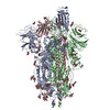
| Title | Structure of a SARS-CoV-2 spike S2 subunit in a pre-fusion, open conformation | |||||||||
 Map data Map data | ||||||||||
 Sample Sample |
| |||||||||
 Keywords Keywords | SARS-CoV-2 / Spike / S2 / VIRAL PROTEIN | |||||||||
| Function / homology |  Function and homology information Function and homology informationvirion component / symbiont-mediated disruption of host tissue / Maturation of spike protein / Translation of Structural Proteins / Virion Assembly and Release / host cell surface / host extracellular region / symbiont-mediated-mediated suppression of host tetherin activity / Induction of Cell-Cell Fusion / structural constituent of virion ...virion component / symbiont-mediated disruption of host tissue / Maturation of spike protein / Translation of Structural Proteins / Virion Assembly and Release / host cell surface / host extracellular region / symbiont-mediated-mediated suppression of host tetherin activity / Induction of Cell-Cell Fusion / structural constituent of virion / positive regulation of viral entry into host cell / membrane fusion / host cell endoplasmic reticulum-Golgi intermediate compartment membrane / Attachment and Entry / entry receptor-mediated virion attachment to host cell / receptor-mediated virion attachment to host cell / host cell surface receptor binding / symbiont-mediated suppression of host innate immune response / endocytosis involved in viral entry into host cell / receptor ligand activity / fusion of virus membrane with host plasma membrane / fusion of virus membrane with host endosome membrane / viral envelope / symbiont entry into host cell / virion attachment to host cell / host cell plasma membrane / SARS-CoV-2 activates/modulates innate and adaptive immune responses / virion membrane / membrane / identical protein binding / plasma membrane Similarity search - Function | |||||||||
| Biological species |   Severe acute respiratory syndrome coronavirus 2 / Severe acute respiratory syndrome coronavirus 2 /  Homo sapiens (human) / Homo sapiens (human) /  Tequatrovirus T4 Tequatrovirus T4 | |||||||||
| Method | single particle reconstruction / cryo EM / Resolution: 2.93 Å | |||||||||
 Authors Authors | Olmedillas E / Diaz R / Hastie K / Ollmann-Saphire E | |||||||||
| Funding support |  United States, 1 items United States, 1 items
| |||||||||
 Citation Citation | Journal: Cell Rep / Year: 2025 Title: Structure of a SARS-CoV-2 spike S2 subunit in a pre-fusion, open conformation. Authors: Eduardo Olmedillas / Roshan R Rajamanickam / Ruben Diaz Avalos / Fernanda Ana-Sosa-Batiz / Dawid Zyla / Michelle A Zandonatti / Stephanie S Harkins / Sujan Shresta / Kathryn M Hastie / Erica Ollmann Saphire / Abstract: The continued emergence of SARS-CoV-2 variants necessitates the development of immunogens that promote broad and durable immunity. The SARS-CoV-2 S2 fusion subunit drives viral entry and has sequence ...The continued emergence of SARS-CoV-2 variants necessitates the development of immunogens that promote broad and durable immunity. The SARS-CoV-2 S2 fusion subunit drives viral entry and has sequence conservation among coronavirus spike proteins. Therefore, S2 could represent an immunogen to boost broadly reactive antibodies. However, when expressed without the S1 domain, metastable S2 irreversibly collapses into the post-fusion six-helix bundle conformation. Beyond well-characterized RBD/NTD shifts, biophysical measurements indicate that spike exhibits reversible "breathing" motions. Using an engineered S2-only antigen that retains the pre-fusion viral surface conformation, we isolated S2-specific antibodies from convalescent and vaccinated individuals. One mAb was used to solve a high-resolution cryo-EM structure of pre-fusion S2. Our structure reveals that, relative to intact spike, engineered S2 adopts a more "open" conformation with stabilizing intermolecular interactions at the trimer base and fusion peptide repositioning. This structure could advance next-generation "booster" immunogens and illuminate potential breathing adjustments of the coronavirus spike. | |||||||||
| History |
|
- Structure visualization
| Supplemental images |
|---|

- Downloads & links
-EMDB archive
| Map data | emd_42985.map.gz | 42 MB | EMDB map data format | |
|---|---|---|---|---|
| Header (meta data) | emd-42985-v30.xmlemd-42985.xml | 26.4 KB 26.4 KB | Display Display | EMDB header |
| Images |  emd_42985.png emd_42985.png | 73.9 KB | ||
| Filedesc metadata | emd-42985.cif.gz | 7.7 KB | ||
| Others | emd_42985_half_map_1.map.gzemd_42985_half_map_2.map.gz | 77.7 MB 77.7 MB | ||
| Archive directory |  http://ftp.pdbj.org/pub/emdb/structures/EMD-42985ftp://ftp.pdbj.org/pub/emdb/structures/EMD-42985 http://ftp.pdbj.org/pub/emdb/structures/EMD-42985ftp://ftp.pdbj.org/pub/emdb/structures/EMD-42985 | HTTPS FTP |
-Related structure data
| Related structure data |  8v5vMC M: atomic model generated by this map C: citing same article ( |
|---|---|
| Similar structure data |
-Links
| EMDB pages | EMDB (EBI/PDBe) / EMDataResource |
|---|---|
| Related items in Molecule of the Month |


-Map
| File | Download / File: emd_42985.map.gz / Format: CCP4 / Size: 83.7 MB / Type: IMAGE STORED AS FLOATING POINT NUMBER (4 BYTES) | ||||||||||||||||||||||||||||||||||||
|---|---|---|---|---|---|---|---|---|---|---|---|---|---|---|---|---|---|---|---|---|---|---|---|---|---|---|---|---|---|---|---|---|---|---|---|---|---|
| Projections & slices | Image control
Images are generated by Spider. | ||||||||||||||||||||||||||||||||||||
| Voxel size | X=Y=Z: 1.32 Å | ||||||||||||||||||||||||||||||||||||
| Density |
| ||||||||||||||||||||||||||||||||||||
| Symmetry | Space group: 1 | ||||||||||||||||||||||||||||||||||||
| Details | EMDB XML:
|
 Z (Sec.)
 Y (Row.)
 X (Col.)


-Supplemental data
-Half map: #1
| File | emd_42985_half_map_1.map | ||||||||||||
|---|---|---|---|---|---|---|---|---|---|---|---|---|---|
| Projections & Slices |
| ||||||||||||
| Density Histograms |


-Half map: #2
| File | emd_42985_half_map_2.map | ||||||||||||
|---|---|---|---|---|---|---|---|---|---|---|---|---|---|
| Projections & Slices |
| ||||||||||||
| Density Histograms |


- Sample components
-Entire : Structure of a SARS-CoV-2 spike S2 subunit in a pre-fusion, open ...
| Entire | Name: Structure of a SARS-CoV-2 spike S2 subunit in a pre-fusion, open conformation in complex with the 6C10 Fab |
|---|---|
| Components |
|
-Supramolecule #1: Structure of a SARS-CoV-2 spike S2 subunit in a pre-fusion, open ...
| Supramolecule | Name: Structure of a SARS-CoV-2 spike S2 subunit in a pre-fusion, open conformation in complex with the 6C10 Fab type: complex / ID: 1 / Parent: 0 / Macromolecule list: #1-#3 |
|---|---|
| Source (natural) | Organism: Severe acute respiratory syndrome coronavirus 2 / Strain: Wuhan |
| Molecular weight | Theoretical: 280 KDa |
-Supramolecule #2: Fab fragment of the 6C10 human monoclonal antibody
| Supramolecule | Name: Fab fragment of the 6C10 human monoclonal antibody / type: complex / ID: 2 / Parent: 1 / Macromolecule list: #2-#3 |
|---|---|
| Source (natural) | Organism: Homo sapiens (human) / Organ: Immune system |
-Supramolecule #3: Structure of a SARS-CoV-2 spike S2 subunit in a pre-fusion, open ...
| Supramolecule | Name: Structure of a SARS-CoV-2 spike S2 subunit in a pre-fusion, open conformation type: complex / ID: 3 / Parent: 1 / Macromolecule list: #1 |
|---|---|
| Source (natural) | Organism: |
-Macromolecule #1: Spike protein S2,Fibritin
| Macromolecule | Name: Spike protein S2,Fibritin / type: protein_or_peptide / ID: 1 / Number of copies: 3 / Enantiomer: LEVO |
|---|---|
| Source (natural) | Organism: Tequatrovirus T4 |
| Molecular weight | Theoretical: 64.292461 KDa |
| Recombinant expression | Organism:  Cricetulus griseus (Chinese hamster) Cricetulus griseus (Chinese hamster) |
| Sequence | String: SIIAYTMSLG AENSVACSNN SIAIPTNFTI SVTTEILPVS MTKTSVDCTM YICGDSTECS NLLLQYGSFC TQLNRALTGI AVEQDKNTQ EVFAQVKQCY KTPPIKDFGG FNFSQILPDP SKPSKRSPIE DLLFNKVTLA DAGFIKQYGD CLGDIAARDL I CAQKFNGL ...String: SIIAYTMSLG AENSVACSNN SIAIPTNFTI SVTTEILPVS MTKTSVDCTM YICGDSTECS NLLLQYGSFC TQLNRALTGI AVEQDKNTQ EVFAQVKQCY KTPPIKDFGG FNFSQILPDP SKPSKRSPIE DLLFNKVTLA DAGFIKQYGD CLGDIAARDL I CAQKFNGL TVLPPLLTDE MIAQYTSCLL AGTICSGWTF GAGPALQIPF PMQMAYRFNG IGVTQNVLYE NQKLIANQFN SA IGKIQDS LSSTPSALGK LQDVVNQNAQ ALNTLVKQLS SNFGAISSVL NDILSRLDKP EAEVQIDRLI TGRLQSLQTY VTQ QLIRAA EIRASANLAA TKMSECVLGQ SKRVDFCGKG YHLMSFPQSA PHGVVFLHVT YVPAQEKNFT TAPAICHDGK AHFP REGVF VSNGTHWFVT QRNFYEPQII TTDNTFVSGN CDVVIGIVNN TVYDPLQPEL DSFKEELDKY FKNHTSPDVD LGDIS GINA SVVNIQKEID RLNEVAKNLN ESLIDLQELG KYEQYIKGSG YIPEAPRDGQ AYVRKDGEWV LLSTFLLEVL FQGPAG WSH PQFEKGGGSG GGSGGGSWSH PQFEK UniProtKB: Spike glycoprotein, Fibritin |
-Macromolecule #2: Variable domain of the light chain from the human antibody 6C10
| Macromolecule | Name: Variable domain of the light chain from the human antibody 6C10 type: protein_or_peptide / ID: 2 / Number of copies: 3 / Enantiomer: LEVO |
|---|---|
| Source (natural) | Organism: Homo sapiens (human) |
| Molecular weight | Theoretical: 11.696993 KDa |
| Recombinant expression | Organism: Cricetulus griseus (Chinese hamster) |
| Sequence | String: AIRMTQSPST LSVSPGERAT LSCRASQSVS SYLAWYQHKP GQAPRLLVYG ASTRATGIPA RFSGSGSGTE FTLTISSLQS EDFAVYYCQ QYNNWPPPFT FGPGTKVDI |
-Macromolecule #3: Variable domain of the heavy chain from the human antibody 6C10
| Macromolecule | Name: Variable domain of the heavy chain from the human antibody 6C10 type: protein_or_peptide / ID: 3 / Number of copies: 3 / Enantiomer: LEVO |
|---|---|
| Source (natural) | Organism: Homo sapiens (human) |
| Molecular weight | Theoretical: 13.913493 KDa |
| Recombinant expression | Organism: Cricetulus griseus (Chinese hamster) |
| Sequence | String: QVQLVQSGAE VRKPGASVKV SCKASGYTFT GYYIHWVRQA PGQGLEWMGW INPNSGGTNY AQKFQGWVIM TRDTSISTAY MELSRLTSD DTAVYYCARC GARSYYYDSG DYCAFDIWGQ GTVVTV |
-Macromolecule #5: 2-acetamido-2-deoxy-beta-D-glucopyranose
| Macromolecule | Name: 2-acetamido-2-deoxy-beta-D-glucopyranose / type: ligand / ID: 5 / Number of copies: 15 / Formula: NAG |
|---|---|
| Molecular weight | Theoretical: 221.208 Da |
| Chemical component information |  ChemComp-NAG: |
-Experimental details
-Structure determination
| Method | cryo EM |
|---|---|
 Processing Processing | single particle reconstruction |
| Aggregation state | particle |
-Sample preparation
| Concentration | 3.0 mg/mL | |||||||||
|---|---|---|---|---|---|---|---|---|---|---|
| Buffer | pH: 8 Component:
Details: Filtered and degased | |||||||||
| Grid | Model: C-flat-2/1 / Material: COPPER / Mesh: 300 / Support film - Material: CARBON / Support film - topology: HOLEY / Support film - Film thickness: 5 / Pretreatment - Type: GLOW DISCHARGE / Pretreatment - Time: 15 sec. / Pretreatment - Atmosphere: AIR / Pretreatment - Pressure: 0.001 kPa | |||||||||
| Vitrification | Cryogen name: ETHANE / Chamber humidity: 100 % / Chamber temperature: 277 K / Instrument: FEI VITROBOT MARK IV / Details: 6 seconds blotting time at force 0. |
- Electron microscopy
| Microscope | FEI TITAN KRIOS |
|---|---|
| Temperature | Min: 80.1 K / Max: 90.2 K |
| Image recording | Film or detector model: GATAN K3 BIOQUANTUM (6k x 4k) / Digitization - Dimensions - Width: 6000 pixel / Digitization - Dimensions - Height: 4000 pixel / Number grids imaged: 2 / Number real images: 12444 / Average exposure time: 1.4 sec. / Average electron dose: 50.0 e/Å2 / Details: Two datasets |
| Electron beam | Acceleration voltage: 300 kV / Electron source:  FIELD EMISSION GUN FIELD EMISSION GUN |
| Electron optics | C2 aperture diameter: 70.0 µm / Calibrated defocus max: 1.973 µm / Calibrated defocus min: 0.481 µm / Calibrated magnification: 75757 / Illumination mode: FLOOD BEAM / Imaging mode: BRIGHT FIELD / Cs: 2.7 mm / Nominal defocus max: 1.8 µm / Nominal defocus min: 0.6 µm / Nominal magnification: 75000 |
| Sample stage | Specimen holder model: FEI TITAN KRIOS AUTOGRID HOLDER / Cooling holder cryogen: NITROGEN |
| Experimental equipment |  Model: Titan Krios / Image courtesy: FEI Company |
+Image processing
-Atomic model buiding 1
| Initial model | PDB ID: Chain - Source name: PDB / Chain - Initial model type: experimental model |
|---|---|
| Refinement | Space: REAL / Protocol: OTHER / Overall B value: 150 / Target criteria: Cross-correlation coefficient |
| Output model | PDB-8v5v: |