Movie
Movie Controller

[English] 日本語
 Yorodumi
- PDB-8v5v: Structure of a SARS-CoV-2 spike S2 subunit in a pre-fusion, open ... -
+ Open data

- Basic information
| Entry | Database: PDB / ID: 8v5v | ||||||
|---|---|---|---|---|---|---|---|
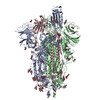
| Title | Structure of a SARS-CoV-2 spike S2 subunit in a pre-fusion, open conformation | ||||||
 Components Components |
| ||||||
 Keywords Keywords | VIRAL PROTEIN / SARS-CoV-2 / Spike / S2 | ||||||
| Function / homology |  Function and homology information Function and homology informationvirion component / symbiont-mediated disruption of host tissue / Maturation of spike protein / Translation of Structural Proteins / Virion Assembly and Release / host cell surface / host extracellular region / symbiont-mediated-mediated suppression of host tetherin activity / Induction of Cell-Cell Fusion / structural constituent of virion ...virion component / symbiont-mediated disruption of host tissue / Maturation of spike protein / Translation of Structural Proteins / Virion Assembly and Release / host cell surface / host extracellular region / symbiont-mediated-mediated suppression of host tetherin activity / Induction of Cell-Cell Fusion / structural constituent of virion / positive regulation of viral entry into host cell / membrane fusion / host cell endoplasmic reticulum-Golgi intermediate compartment membrane / Attachment and Entry / entry receptor-mediated virion attachment to host cell / receptor-mediated virion attachment to host cell / host cell surface receptor binding / symbiont-mediated suppression of host innate immune response / endocytosis involved in viral entry into host cell / receptor ligand activity / fusion of virus membrane with host plasma membrane / fusion of virus membrane with host endosome membrane / viral envelope / symbiont entry into host cell / virion attachment to host cell / host cell plasma membrane / SARS-CoV-2 activates/modulates innate and adaptive immune responses / virion membrane / membrane / identical protein binding / plasma membrane Similarity search - Function | ||||||
| Biological species |   Severe acute respiratory syndrome coronavirus 2Tequatrovirus T4 Severe acute respiratory syndrome coronavirus 2Tequatrovirus T4 Homo sapiens (human) Homo sapiens (human) | ||||||
| Method | ELECTRON MICROSCOPY / single particle reconstruction / cryo EM / Resolution: 2.93 Å | ||||||
 Authors Authors | Olmedillas, E. / Diaz, R. / Hastie, K. / Ollmann-Saphire, E. | ||||||
| Funding support |  United States, 1items United States, 1items
| ||||||
 Citation Citation | Journal: Cell Rep / Year: 2025 Title: Structure of a SARS-CoV-2 spike S2 subunit in a pre-fusion, open conformation. Authors: Eduardo Olmedillas / Roshan R Rajamanickam / Ruben Diaz Avalos / Fernanda Ana-Sosa-Batiz / Dawid Zyla / Michelle A Zandonatti / Stephanie S Harkins / Sujan Shresta / Kathryn M Hastie / Erica Ollmann Saphire / Abstract: The continued emergence of SARS-CoV-2 variants necessitates the development of immunogens that promote broad and durable immunity. The SARS-CoV-2 S2 fusion subunit drives viral entry and has sequence ...The continued emergence of SARS-CoV-2 variants necessitates the development of immunogens that promote broad and durable immunity. The SARS-CoV-2 S2 fusion subunit drives viral entry and has sequence conservation among coronavirus spike proteins. Therefore, S2 could represent an immunogen to boost broadly reactive antibodies. However, when expressed without the S1 domain, metastable S2 irreversibly collapses into the post-fusion six-helix bundle conformation. Beyond well-characterized RBD/NTD shifts, biophysical measurements indicate that spike exhibits reversible "breathing" motions. Using an engineered S2-only antigen that retains the pre-fusion viral surface conformation, we isolated S2-specific antibodies from convalescent and vaccinated individuals. One mAb was used to solve a high-resolution cryo-EM structure of pre-fusion S2. Our structure reveals that, relative to intact spike, engineered S2 adopts a more "open" conformation with stabilizing intermolecular interactions at the trimer base and fusion peptide repositioning. This structure could advance next-generation "booster" immunogens and illuminate potential breathing adjustments of the coronavirus spike. | ||||||
| History |
|
- Structure visualization
| Structure viewer | Molecule: MolmilJmol/JSmol |
|---|
- Downloads & links
-Download
| PDBx/mmCIF format | 8v5v.cif.gz | 405.1 KB | Display | PDBx/mmCIF format |
|---|---|---|---|---|
| PDB format | pdb8v5v.ent.gz | 326.8 KB | Display | PDB format |
| PDBx/mmJSON format | 8v5v.json.gz | Tree view | PDBx/mmJSON format | |
| Others |  Other downloads Other downloads |
-Validation report
| Arichive directory | https://data.pdbj.org/pub/pdb/validation_reports/v5/8v5vftp://data.pdbj.org/pub/pdb/validation_reports/v5/8v5v | HTTPS FTP |
|---|
-Related structure data
| Related structure data |  42985MC M: map data used to model this data C: citing same article ( |
|---|---|
| Similar structure data |
-Links
 PDBj
PDBj


- Assembly
| Deposited unit | 
|
|---|---|
| 1 |
|
-Components
| #1: Protein | Mass: 64292.461 Da / Num. of mol.: 3 Mutation: F817P,A892P,A899P,A942P,V987P,Y707C,T883C,Y788C,A876C Source method: isolated from a genetically manipulated source Source: (gene. exp.) Severe acute respiratory syndrome coronavirus 2, (gene. exp.) Tequatrovirus T4Gene: S, 2, wac / Production host:   Cricetulus griseus (Chinese hamster) / References: UniProt: P0DTC2, UniProt: P10104 Cricetulus griseus (Chinese hamster) / References: UniProt: P0DTC2, UniProt: P10104#2: Antibody | Mass: 11696.993 Da / Num. of mol.: 3 Source method: isolated from a genetically manipulated source Source: (gene. exp.) Homo sapiens (human) / Production host: Cricetulus griseus (Chinese hamster)#3: Antibody | Mass: 13913.493 Da / Num. of mol.: 3 Source method: isolated from a genetically manipulated source Source: (gene. exp.) Homo sapiens (human) / Production host: Cricetulus griseus (Chinese hamster)#4: Polysaccharide | Source method: isolated from a genetically manipulated source #5: Sugar | ChemComp-NAG /   Type: D-saccharide, beta linking / Mass: 221.208 Da / Num. of mol.: 15 / Source method: obtained synthetically / Formula: C8H15NO6 Type: D-saccharide, beta linking / Mass: 221.208 Da / Num. of mol.: 15 / Source method: obtained synthetically / Formula: C8H15NO6Has ligand of interest | N | Has protein modification | Y | |
|---|
-Experimental details
-Experiment
| Experiment | Method: ELECTRON MICROSCOPY |
|---|---|
| EM experiment | Aggregation state: PARTICLE / 3D reconstruction method: single particle reconstruction |
- Sample preparation
| Component |
| ||||||||||||||||||||||||
|---|---|---|---|---|---|---|---|---|---|---|---|---|---|---|---|---|---|---|---|---|---|---|---|---|---|
| Molecular weight |
| ||||||||||||||||||||||||
| Source (natural) |
| ||||||||||||||||||||||||
| Source (recombinant) |
| ||||||||||||||||||||||||
| Buffer solution | pH: 8 / Details: Filtered and degased | ||||||||||||||||||||||||
| Buffer component |
| ||||||||||||||||||||||||
| Specimen | Conc.: 3 mg/ml / Embedding applied: NO / Shadowing applied: NO / Staining applied: NO / Vitrification applied: YES | ||||||||||||||||||||||||
| Specimen support | Grid material: COPPER / Grid mesh size: 300 divisions/in. / Grid type: C-flat-2/1 | ||||||||||||||||||||||||
| Vitrification | Instrument: FEI VITROBOT MARK IV / Cryogen name: ETHANE / Humidity: 100 % / Chamber temperature: 277 K / Details: 6 seconds blotting time at force 0 |
- Electron microscopy imaging
| Experimental equipment |  Model: Titan Krios / Image courtesy: FEI Company |
|---|---|
| Microscopy | Model: FEI TITAN KRIOS |
| Electron gun | Electron source:  FIELD EMISSION GUN / Accelerating voltage: 300 kV / Illumination mode: FLOOD BEAM FIELD EMISSION GUN / Accelerating voltage: 300 kV / Illumination mode: FLOOD BEAM |
| Electron lens | Mode: BRIGHT FIELD / Nominal magnification: 75000 X / Calibrated magnification: 75757 X / Nominal defocus max: 1800 nm / Nominal defocus min: 600 nm / Calibrated defocus min: 481 nm / Calibrated defocus max: 1973 nm / Cs: 2.7 mm / C2 aperture diameter: 70 µm / Alignment procedure: ZEMLIN TABLEAU |
| Specimen holder | Cryogen: NITROGEN / Specimen holder model: FEI TITAN KRIOS AUTOGRID HOLDER / Temperature (max): 90.2 K / Temperature (min): 80.1 K / Residual tilt: 0.1 mradians |
| Image recording | Average exposure time: 1.4 sec. / Electron dose: 50 e/Å2 / Film or detector model: GATAN K3 BIOQUANTUM (6k x 4k) / Num. of grids imaged: 2 / Num. of real images: 12444 / Details: Two datasets |
| Image scans | Sampling size: 5 µm / Width: 6000 / Height: 4000 |
- Processing
| EM software |
| ||||||||||||||||||||||||||||||||||||||||||||||||||
|---|---|---|---|---|---|---|---|---|---|---|---|---|---|---|---|---|---|---|---|---|---|---|---|---|---|---|---|---|---|---|---|---|---|---|---|---|---|---|---|---|---|---|---|---|---|---|---|---|---|---|---|
| CTF correction | Type: PHASE FLIPPING AND AMPLITUDE CORRECTION | ||||||||||||||||||||||||||||||||||||||||||||||||||
| Particle selection | Num. of particles selected: 407314 | ||||||||||||||||||||||||||||||||||||||||||||||||||
| Symmetry | Point symmetry: C3 (3 fold cyclic) | ||||||||||||||||||||||||||||||||||||||||||||||||||
| 3D reconstruction | Resolution: 2.93 Å / Resolution method: FSC 0.143 CUT-OFF / Num. of particles: 408064 / Algorithm: FOURIER SPACE / Num. of class averages: 1 / Symmetry type: POINT | ||||||||||||||||||||||||||||||||||||||||||||||||||
| Atomic model building |
| ||||||||||||||||||||||||||||||||||||||||||||||||||
| Atomic model building |
| ||||||||||||||||||||||||||||||||||||||||||||||||||
| Refine LS restraints |
|