Movie
Movie Controller

[English] 日本語
 Yorodumi
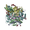
- PDB-9dtu: Structure of the Measles virus Fusion protein ectodomain E170G E4... -
+ Open data

- Basic information
| Entry | Database: PDB / ID: 9dtu | ||||||||||||||||||||||||||||||
|---|---|---|---|---|---|---|---|---|---|---|---|---|---|---|---|---|---|---|---|---|---|---|---|---|---|---|---|---|---|---|---|
| Title | Structure of the Measles virus Fusion protein ectodomain E170G E455G with mutated furin cleavage site in the pre-fusion conformation | ||||||||||||||||||||||||||||||
 Components Components | Fusion glycoprotein F0 | ||||||||||||||||||||||||||||||
 Keywords Keywords | VIRAL PROTEIN / glycoprotein / immune system / measles / high-resolution / ectodomain / pre-fusion | ||||||||||||||||||||||||||||||
| Function / homology | Precursor fusion glycoprotein F0, Paramyxoviridae / Fusion glycoprotein F0 / fusion of virus membrane with host plasma membrane / viral envelope / symbiont entry into host cell / host cell plasma membrane / virion membrane / Fusion glycoprotein F0 Function and homology information Function and homology information | ||||||||||||||||||||||||||||||
| Biological species |  Measles virus strain Ichinose-B95a Measles virus strain Ichinose-B95a | ||||||||||||||||||||||||||||||
| Method | ELECTRON MICROSCOPY / single particle reconstruction / cryo EM / Resolution: 2.05 Å | ||||||||||||||||||||||||||||||
 Authors Authors | Zyla, D. / Saphire, E.O. | ||||||||||||||||||||||||||||||
| Funding support |  Switzerland, Switzerland,  United States, 5items United States, 5items
| ||||||||||||||||||||||||||||||
 Citation Citation | Journal: To Be Published Title: Full-length soluble measles fusion glycoprotein as a new subunit vaccine strategy Authors: Zyla, D. / Saphire, E.O. | ||||||||||||||||||||||||||||||
| History |
|
- Structure visualization
| Structure viewer | Molecule: MolmilJmol/JSmol |
|---|
- Downloads & links
-Download
| PDBx/mmCIF format | 9dtu.cif.gz | 339.6 KB | Display | PDBx/mmCIF format |
|---|---|---|---|---|
| PDB format | pdb9dtu.ent.gz | 220.5 KB | Display | PDB format |
| PDBx/mmJSON format | 9dtu.json.gz | Tree view | PDBx/mmJSON format | |
| Others |  Other downloads Other downloads |
-Validation report
| Arichive directory | https://data.pdbj.org/pub/pdb/validation_reports/dt/9dtuftp://data.pdbj.org/pub/pdb/validation_reports/dt/9dtu | HTTPS FTP |
|---|
-Related structure data
| Related structure data |  47166MC  9coeC  9cofC  9cogC  9cohC M: map data used to model this data C: citing same article ( |
|---|---|
| Similar structure data |


-Links
 PDBj
PDBj

- Assembly
| Deposited unit | 
|
|---|---|
| 1 |
|
-Components
| #1: Protein | Mass: 56819.855 Da / Num. of mol.: 3 Source method: isolated from a genetically manipulated source Source: (gene. exp.) Measles virus strain Ichinose-B95a / Strain: Ichinose-B95a / Cell line (production host): ExpiCHO / Production host:   Cricetulus griseus (Chinese hamster) / References: UniProt: Q786F3 Cricetulus griseus (Chinese hamster) / References: UniProt: Q786F3#2: Polysaccharide | 2-acetamido-2-deoxy-beta-D-glucopyranose-(1-4)-2-acetamido-2-deoxy-beta-D-glucopyranose Source method: isolated from a genetically manipulated source #3: Sugar |   Type: D-saccharide, beta linking / Mass: 221.208 Da / Num. of mol.: 3 / Source method: obtained synthetically / Formula: C8H15NO6 Type: D-saccharide, beta linking / Mass: 221.208 Da / Num. of mol.: 3 / Source method: obtained synthetically / Formula: C8H15NO6#4: Water | ChemComp-HOH / |  Mass: 18.015 Da / Num. of mol.: 400 / Source method: isolated from a natural source / Formula: H2O Mass: 18.015 Da / Num. of mol.: 400 / Source method: isolated from a natural source / Formula: H2OHas ligand of interest | N | Has protein modification | Y | |
|---|
-Experimental details
-Experiment
| Experiment | Method: ELECTRON MICROSCOPY |
|---|---|
| EM experiment | Aggregation state: PARTICLE / 3D reconstruction method: single particle reconstruction |
- Sample preparation
| Component | Name: Structure of the Measles virus Fusion protein ectodomain E170G E455G with mutated furin cleavage site in the pre-fusion conformation Type: COMPLEX / Entity ID: #1 / Source: RECOMBINANT |
|---|---|
| Molecular weight | Value: 0.15 MDa / Experimental value: NO |
| Source (natural) | Organism:  Measles morbillivirus / Strain: Ichinose-B95a Measles morbillivirus / Strain: Ichinose-B95a |
| Source (recombinant) | Organism: Cricetulus griseus (Chinese hamster) / Strain: ExpiCHO |
| Buffer solution | pH: 8 / Details: HEPES 50 mM, pH 8.0, NaCl 150 mM |
| Specimen | Conc.: 0.2 mg/ml / Embedding applied: NO / Shadowing applied: NO / Staining applied: NO / Vitrification applied: YES |
| Specimen support | Grid material: COPPER / Grid mesh size: 300 divisions/in. / Grid type: Quantifoil |
| Vitrification | Instrument: FEI VITROBOT MARK IV / Cryogen name: ETHANE / Humidity: 100 % / Chamber temperature: 298 K |
- Electron microscopy imaging
| Experimental equipment |  Model: Titan Krios / Image courtesy: FEI Company |
|---|---|
| Microscopy | Model: TFS KRIOS |
| Electron gun | Electron source:  FIELD EMISSION GUN / Accelerating voltage: 300 kV / Illumination mode: FLOOD BEAM FIELD EMISSION GUN / Accelerating voltage: 300 kV / Illumination mode: FLOOD BEAM |
| Electron lens | Mode: BRIGHT FIELD / Nominal defocus max: 2500 nm / Nominal defocus min: 1000 nm |
| Specimen holder | Cryogen: NITROGEN / Specimen holder model: FEI TITAN KRIOS AUTOGRID HOLDER |
| Image recording | Average exposure time: 2 sec. / Electron dose: 70 e/Å2 / Film or detector model: GATAN K3 (6k x 4k) / Num. of grids imaged: 3 / Num. of real images: 10974 |
- Processing
| EM software | Name: PHENIX / Version: 1.21_5184 / Category: model refinement | ||||||||||||||||||||||||
|---|---|---|---|---|---|---|---|---|---|---|---|---|---|---|---|---|---|---|---|---|---|---|---|---|---|
| CTF correction | Type: PHASE FLIPPING AND AMPLITUDE CORRECTION | ||||||||||||||||||||||||
| Particle selection | Num. of particles selected: 1500000 | ||||||||||||||||||||||||
| 3D reconstruction | Resolution: 2.05 Å / Resolution method: FSC 0.143 CUT-OFF / Num. of particles: 281000 / Symmetry type: POINT | ||||||||||||||||||||||||
| Atomic model building | Protocol: RIGID BODY FIT / Space: REAL | ||||||||||||||||||||||||
| Atomic model building | PDB-ID: 8UUP Accession code: 8UUP / Source name: PDB / Type: experimental model | ||||||||||||||||||||||||
| Refinement | Cross valid method: NONE Stereochemistry target values: GeoStd + Monomer Library + CDL v1.2 | ||||||||||||||||||||||||
| Displacement parameters | Biso mean: 27.3 Å2 | ||||||||||||||||||||||||
| Refine LS restraints |
|