 ムービー
ムービー コントローラー
 構造ビューア
 EMN検索について


-検索条件
-検索結果
検索 (著者・登録者: savva & c)の結果119件中、1から50件目までを表示しています

EMDB-18290: 
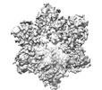
Cryo-EM structure of Cx26 gap junction K125E mutant in bicarbonate buffer (classification on hemichannel)

EMDB-18291:
Cryo-EM structure of Cx26 solubilised in LMNG - hemichannel classification - NConst conformation

EMDB-18292:
Cryo-EM structure of Cx26 solubilised in LMNG - Hemichannel classification NFlex conformation
EMDB-18293:
Cryo-EM structure of Cx26 solubilised in LMNG: classification on subunit A; Nconst-mon conformation

EMDB-18294:
Cryo-EM structure of Cx26 solubilised in LMNG: classification on subunit A; NFlex conformation

EMDB-18295:
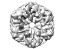
Cryo-EM reconstruction of Cx26 gap junction K125R mutant (D6 symmetry)
EMDB-18296:
Cryo-EM reconstruction of Cx26 gap junction K125E mutant in HEPES buffer

EMDB-18297:
Cryo-EM reconstruction of Cx26 gap junction WT in HEPES buffer

PDB-8q9z:
Cryo-EM structure of Cx26 gap junction K125E mutant in bicarbonate buffer (classification on hemichannel)

PDB-8qa0:
Cryo-EM structure of Cx26 solubilised in LMNG - hemichannel classification - NConst conformation

PDB-8qa1:
Cryo-EM structure of Cx26 solubilised in LMNG - Hemichannel classification NFlex conformation

PDB-8qa2:
Cryo-EM structure of Cx26 solubilised in LMNG: classification on subunit A; Nconst-mon conformation

PDB-8qa3:
Cryo-EM structure of Cx26 solubilised in LMNG: classification on subunit A; NFlex conformation

EMDB-50185:
KS + AT di-domain of polyketide synthase 13 in Mycobacterium tuberculosis

EMDB-39478:
Structure of Aquifex aeolicus Lumazine Synthase by Cryo-Electron Microscopy to 1.42 Angstrom Resolution

PDB-8yt4:
Structure of Aquifex aeolicus Lumazine Synthase by Cryo-Electron Microscopy to 1.42 Angstrom Resolution

EMDB-16820:
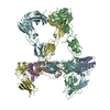
Cryo-EM structure of a pre-dimerized murine IL-12 complete extracellular signaling complex (Class 1).

EMDB-16821:
Cryo-EM structure of a pre-dimerized murine IL-12 complete extracellular signaling complex (Class 2).

EMDB-16822:
Cryo-EM structure of the murine IL-12 complete extracellular signaling complex (Class 1).

EMDB-16823:
Cryo-EM structure of the murine IL-12 complete extracellular signaling complex (Class 2).

EMDB-16824:
Cryo-EM structure of a pre-dimerized human IL-23 complete extracellular signaling complex.

EMDB-17580:
Cryo-EM structure of a pre-dimerized murine IL-12 complete extracellular signaling complex (Class 1), obtained after local refinement.

PDB-8odz:
Cryo-EM structure of a pre-dimerized murine IL-12 complete extracellular signaling complex (Class 1).

PDB-8oe0:
Cryo-EM structure of a pre-dimerized murine IL-12 complete extracellular signaling complex (Class 2).

PDB-8oe4:
Cryo-EM structure of a pre-dimerized human IL-23 complete extracellular signaling complex.

PDB-8pb1:
Cryo-EM structure of a pre-dimerized murine IL-12 complete extracellular signaling complex (Class 1), obtained after local refinement.

EMDB-15677:
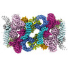
Cryo-EM structure for mouse leptin in complex with the mouse LEP-R ectodomain (1:2 mLEP:mLEPR model)

EMDB-15678:
Mouse leptin:LEP-R complex cryoEM structure (3:3 model)

EMDB-15679:
Cryo-EM structure for a 3:3 complex between mouse leptin and the mouse LEP-R ectodomain (local refinement)

EMDB-15680:
Human leptin in complex with the human LEP-R ectodomain fused to a C-terminal trimeric isoleucine GCN4 zipper (2:2 model)

EMDB-15681:
Human leptin in complex with the human LEP-R ectodomain fused to a C-terminal trimeric isoleucine GCN4 zipper (closed 3:3 model)

EMDB-15683:
Human leptin in complex with the human LEP-R ectodomain fused to a C-terminal trimeric isoleucine GCN4 zipper (open 3:3 model).

EMDB-15899:
Cryo-EM structure for the mouse LEPR-CRH2:Leptin:LEPR-Ig complex following symmetry expansion in combination with local refinement

PDB-8avb:
Cryo-EM structure for mouse leptin in complex with the mouse LEP-R ectodomain (1:2 mLEP:mLEPR model).

PDB-8avc:
Mouse leptin:LEP-R complex cryoEM structure (3:3 model)

PDB-8avd:
Cryo-EM structure for a 3:3 complex between mouse leptin and the mouse LEP-R ectodomain (local refinement)

PDB-8ave:
Human leptin in complex with the human LEP-R ectodomain fused to a C-terminal trimeric isoleucine GCN4 zipper (2:2 model)
PDB-8avf:
Human leptin in complex with the human LEP-R ectodomain fused to a C-terminal trimeric isoleucine GCN4 zipper (closed 3:3 model)

PDB-8avo:
Human leptin in complex with the human LEP-R ectodomain fused to a C-terminal trimeric isoleucine GCN4 zipper (open 3:3 model).

PDB-8b7q:
Cryo-EM structure for the mouse LEPR-CRH2:Leptin:LEPR-Ig complex following symmetry expansion in combination with local refinement
EMDB-13594:
Cryo-EM structure of Saccharomyces cerevisiae TOROID (TORC1 Organized in Inhibited Domains).

EMDB-13595:
Helical reconstruction of TOROID (TORC1 Organized in Inhibited Domains) filaments.
PDB-7pqh:
Cryo-EM structure of Saccharomyces cerevisiae TOROID (TORC1 Organized in Inhibited Domains).

EMDB-14971:
Cryo-EM structure of the indirubin-bound Hsp90-XAP2-AHR complex

EMDB-14972:
Cryo-EM structure of the indirubin-bound Hsp90-XAP2-AHR complex (focused map).
PDB-7zub:
Cryo-EM structure of the indirubin-bound Hsp90-XAP2-AHR complex

EMDB-13664:
GA1 bacteriophage portal protein

EMDB-13665:
PhiCPV4 bacteriophage Portal Protein

PDB-7pv2:
GA1 bacteriophage portal protein

PDB-7pv4:
PhiCPV4 bacteriophage Portal Protein
ページ:
 wwPDBはEMDBデータモデルのバージョン3へ移行します
