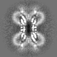
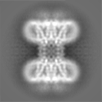
- EMDB-18297: Cryo-EM reconstruction of Cx26 gap junction WT in HEPES buffer -
+
Open data
ID or keywords:
Loading...
-
Basic information
Entry
Database: EMDB / ID: EMD-18297
Title
Cryo-EM reconstruction of Cx26 gap junction WT in HEPES buffer
Map data
Full map D6 averaged (unsharpened)
Sample
Complex: Connexin 26 WT in HEPES buffer pH 8.0
Protein or peptide: Connexin 26
Keywords
Gap junction Large Pore Channel Carbon dioxide sensitive / MEMBRANE PROTEIN
Function / homology
Function and homology information
Transport of connexons to the plasma membrane / gap junction channel activity involved in cell communication by electrical coupling / gap junction-mediated intercellular transport / Oligomerization of connexins into connexons / Transport of connexins along the secretory pathway / gap junction assembly / connexin complex / gap junction / Gap junction assembly / gap junction channel activity ...Transport of connexons to the plasma membrane / gap junction channel activity involved in cell communication by electrical coupling / gap junction-mediated intercellular transport / Oligomerization of connexins into connexons / Transport of connexins along the secretory pathway / gap junction assembly / connexin complex / gap junction / Gap junction assembly / gap junction channel activity / endoplasmic reticulum-Golgi intermediate compartment / sensory perception of sound / transmembrane transport / cell-cell signaling / calcium ion binding / identical protein binding / plasma membrane Similarity search - Function
Gap junction beta-2 protein (Cx26) / Connexin / Connexin, N-terminal / Connexin, conserved site / Gap junction protein, cysteine-rich domain / Connexin, N-terminal domain superfamily / Connexin / Connexins signature 1. / Connexins signature 2. / Connexin homologues / Gap junction channel protein cysteine-rich domain Similarity search - Domain/homology
Journal: Elife / Year: 2024 Title: Structures of wild-type and a constitutively closed mutant of connexin26 shed light on channel regulation by CO. Authors: Deborah H Brotherton / Sarbjit Nijjar / Christos G Savva / Nicholas Dale / Alexander David Cameron / Abstract: Connexins allow intercellular communication by forming gap junction channels (GJCs) between juxtaposed cells. Connexin26 (Cx26) can be regulated directly by CO. This is proposed to be mediated ...Connexins allow intercellular communication by forming gap junction channels (GJCs) between juxtaposed cells. Connexin26 (Cx26) can be regulated directly by CO. This is proposed to be mediated through carbamylation of K125. We show that mutating K125 to glutamate, mimicking the negative charge of carbamylation, causes Cx26 GJCs to be constitutively closed. Through cryo-EM we observe that the K125E mutation pushes a conformational equilibrium towards the channel having a constricted pore entrance, similar to effects seen on raising the partial pressure of CO. In previous structures of connexins, the cytoplasmic loop, important in regulation and where K125 is located, is disordered. Through further cryo-EM studies we trap distinct states of Cx26 and observe density for the cytoplasmic loop. The interplay between the position of this loop, the conformations of the transmembrane helices and the position of the N-terminal helix, which controls the aperture to the pore, provides a mechanism for regulation.
Details: The buffer, except DDM and DTT, was prepared fresh from 10x stock on day of used. The basal buffer was filtered and degassed and DDM and DTT added.
Vitrification
Cryogen name: ETHANE-PROPANE / Instrument: LEICA EM GP Details: 3 microlitres protein applied to grid, blot time 3 seconds.
-
Electron microscopy
Microscope
FEI TITAN KRIOS
Specialist optics
Phase plate: VOLTA PHASE PLATE / Energy filter - Name: GIF Bioquantum
Image recording
Film or detector model: FEI FALCON III (4k x 4k) / Detector mode: COUNTING / Number real images: 2436 / Average exposure time: 60.0 sec. / Average electron dose: 39.0 e/Å2
Electron beam
Acceleration voltage: 300 kV / Electron source: FIELD EMISSION GUN
In the structure databanks used in Yorodumi, some data are registered as the other names, "COVID-19 virus" and "2019-nCoV". Here are the details of the virus and the list of structure data.
Jan 31, 2019. EMDB accession codes are about to change! (news from PDBe EMDB page)
EMDB accession codes are about to change! (news from PDBe EMDB page)
The allocation of 4 digits for EMDB accession codes will soon come to an end. Whilst these codes will remain in use, new EMDB accession codes will include an additional digit and will expand incrementally as the available range of codes is exhausted. The current 4-digit format prefixed with “EMD-” (i.e. EMD-XXXX) will advance to a 5-digit format (i.e. EMD-XXXXX), and so on. It is currently estimated that the 4-digit codes will be depleted around Spring 2019, at which point the 5-digit format will come into force.
The EM Navigator/Yorodumi systems omit the EMD- prefix.
Related info.:Q: What is EMD? / ID/Accession-code notation in Yorodumi/EM Navigator
Yorodumi is a browser for structure data from EMDB, PDB, SASBDB, etc.
This page is also the successor to EM Navigator detail page, and also detail information page/front-end page for Omokage search.
The word "yorodu" (or yorozu) is an old Japanese word meaning "ten thousand". "mi" (miru) is to see.
Related info.:EMDB / PDB / SASBDB / Comparison of 3 databanks / Yorodumi Search / Aug 31, 2016. New EM Navigator & Yorodumi / Yorodumi Papers / Jmol/JSmol / Function and homology information / Changes in new EM Navigator and Yorodumi
 Movie
Movie Controller


 Open data
 Basic information
 Map data
 Sample
 Keywords
 Function and homology information
 Homo sapiens (human)
 Authors
 United Kingdom, 2 items
  Citation
 Structure visualization
 Downloads & links
 emd_18297.png
 http://ftp.pdbj.org/pub/emdb/structures/EMD-18297


 Z (Sec.)
 Y (Row.)
 X (Col.)


 Sample components
 Spodoptera frugiperda (fall armyworm)
 Processing
 Electron microscopy
 FIELD EMISSION GUN


