Movie
Movie Controller


+ Open data

- Basic information
| Entry | Database: PDB / ID: 7pv2 | |||||||||||||||
|---|---|---|---|---|---|---|---|---|---|---|---|---|---|---|---|---|
| Title | GA1 bacteriophage portal protein | |||||||||||||||
 Components Components | Head-tail connector (Portal protein) | |||||||||||||||
 Keywords Keywords | VIRAL PROTEIN / Bacteriophage / Portal protein / cryoEM | |||||||||||||||
| Function / homology | Portal protein Gp10 / Portal protein Gp10 superfamily / Phage Connector (GP10) / Head-tail connector (Portal protein) Function and homology information Function and homology information | |||||||||||||||
| Biological species |  Bacillus phage GA-1 (virus) Bacillus phage GA-1 (virus) | |||||||||||||||
| Method | ELECTRON MICROSCOPY / single particle reconstruction / cryo EM / Resolution: 3.2 Å | |||||||||||||||
 Authors Authors | Javed, A. / Villanueva, H. / Orlova, E.V. / Savva, R. | |||||||||||||||
| Funding support |  United Kingdom, 4items United Kingdom, 4items
| |||||||||||||||
 Citation Citation | Journal: Viruses / Year: 2021 Title: Cryo-EM Structures of Two Bacteriophage Portal Proteins Provide Insights for Antimicrobial Phage Engineering. Authors: Abid Javed / Hugo Villanueva / Shadikejiang Shataer / Sara Vasciaveo / Renos Savva / Elena V Orlova / Abstract: Widespread antibiotic resistance has returned attention to bacteriophages as a means of managing bacterial pathogenesis. Synthetic biology approaches to engineer phages have demonstrated genomic ...Widespread antibiotic resistance has returned attention to bacteriophages as a means of managing bacterial pathogenesis. Synthetic biology approaches to engineer phages have demonstrated genomic editing to broaden natural host ranges, or to optimise microbicidal action. Gram positive pathogens cause serious pastoral animal and human infections that are especially lethal in newborns. Such pathogens are targeted by the obligate lytic phages of the and families. These phages have relatively small ~20 kb linear protein-capped genomes and their compact organisation, relatively few structural elements, and broad host range, are appealing from a phage-engineering standpoint. In this study, we focus on portal proteins, which are core elements for the assembly of such tailed phages. The structures of dodecameric portal complexes from phage GA1, which targets , and phage phiCPV4 that infects , were determined at resolutions of 3.3 Å and 2.9 Å, respectively. Both are found to closely resemble the related phi29 portal protein fold. However, the portal protein of phiCPV4 exhibits interesting differences in the clip domain. These structures provide new insights on structural diversity in portal proteins and will be essential for considerations in phage structural engineering. | |||||||||||||||
| History |
|
- Structure visualization
| Structure viewer | Molecule: MolmilJmol/JSmol |
|---|
- Downloads & links
-Download
| PDBx/mmCIF format | 7pv2.cif.gz | 543.5 KB | Display | PDBx/mmCIF format |
|---|---|---|---|---|
| PDB format | pdb7pv2.ent.gz | 456 KB | Display | PDB format |
| PDBx/mmJSON format | 7pv2.json.gz | Tree view | PDBx/mmJSON format | |
| Others |  Other downloads Other downloads |
-Validation report
| Arichive directory | https://data.pdbj.org/pub/pdb/validation_reports/pv/7pv2ftp://data.pdbj.org/pub/pdb/validation_reports/pv/7pv2 | HTTPS FTP |
|---|
-Related structure data
| Related structure data |  13664MC  7pv4C M: map data used to model this data C: citing same article ( |
|---|---|
| Similar structure data |

-Links
 PDBj
PDBj- Assembly
| Deposited unit | 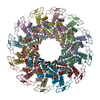
|
|---|---|
| 1 |
|
-Components
| #1: Protein | Mass: 35307.801 Da / Num. of mol.: 12 Source method: isolated from a genetically manipulated source Details: In each subunit of the dodecamer, the DNA Tunnel loop residues (232 - 250) has not been modelled due to lack of EM density. Also missing in the pdb structure are 22 residues at the C- ...Details: In each subunit of the dodecamer, the DNA Tunnel loop residues (232 - 250) has not been modelled due to lack of EM density. Also missing in the pdb structure are 22 residues at the C-terminus and 7 residues at the N-terminus in each chain. Source: (gene. exp.) Bacillus phage GA-1 (virus) / Gene: gene 10 / Production host:  |
|---|
-Experimental details
-Experiment
| Experiment | Method: ELECTRON MICROSCOPY |
|---|---|
| EM experiment | Aggregation state: PARTICLE / 3D reconstruction method: single particle reconstruction |
- Sample preparation
| Component | Name: GA1 bacteriophage portal protein / Type: COMPLEX / Details: A dodecamer complex made up of 12 subunits. / Entity ID: all / Source: RECOMBINANT |
|---|---|
| Source (natural) | Organism: Bacillus phage GA-1 (virus) |
| Source (recombinant) | Organism: |
| Natural host | Organism: Bacillus pumilus |
| Buffer solution | pH: 7.5 |
| Specimen | Conc.: 0.05 mg/ml / Embedding applied: NO / Shadowing applied: NO / Staining applied: NO / Vitrification applied: YES |
| Specimen support | Grid material: COPPER / Grid type: PELCO Ultrathin Carbon with Lacey Carbon |
| Vitrification | Instrument: FEI VITROBOT MARK IV / Cryogen name: ETHANE / Humidity: 100 % / Chamber temperature: 281 K / Details: 3ul of sample was applied on lacey carbon grids. |
- Electron microscopy imaging
| Experimental equipment |  Model: Titan Krios / Image courtesy: FEI Company |
|---|---|
| Microscopy | Model: FEI TITAN KRIOS |
| Electron gun | Electron source:  FIELD EMISSION GUN / Accelerating voltage: 300 kV / Illumination mode: FLOOD BEAM FIELD EMISSION GUN / Accelerating voltage: 300 kV / Illumination mode: FLOOD BEAM |
| Electron lens | Mode: BRIGHT FIELD / Nominal defocus max: 2000 nm / Nominal defocus min: 1200 nm / Cs: 2.7 mm / C2 aperture diameter: 70 µm / Alignment procedure: ZEMLIN TABLEAU |
| Specimen holder | Cryogen: NITROGEN / Specimen holder model: FEI TITAN KRIOS AUTOGRID HOLDER / Temperature (max): 98 K / Temperature (min): 95 K |
| Image recording | Electron dose: 1 e/Å2 / Detector mode: COUNTING / Film or detector model: GATAN K2 SUMMIT (4k x 4k) / Num. of grids imaged: 1 / Num. of real images: 2249 |
| EM imaging optics | Energyfilter name: GIF Quantum LS / Energyfilter slit width: 20 eV |
| Image scans | Sampling size: 5 µm / Width: 3838 / Height: 3710 / Used frames/image: 1-12 |
- Processing
| Software | Name: PHENIX / Version: 1.15.1_3469: / Classification: refinement | ||||||||||||||||||||||||||||||||||||||||||||||||
|---|---|---|---|---|---|---|---|---|---|---|---|---|---|---|---|---|---|---|---|---|---|---|---|---|---|---|---|---|---|---|---|---|---|---|---|---|---|---|---|---|---|---|---|---|---|---|---|---|---|
| EM software |
| ||||||||||||||||||||||||||||||||||||||||||||||||
| CTF correction | Type: PHASE FLIPPING AND AMPLITUDE CORRECTION | ||||||||||||||||||||||||||||||||||||||||||||||||
| Particle selection | Num. of particles selected: 50466 | ||||||||||||||||||||||||||||||||||||||||||||||||
| Symmetry | Point symmetry: C12 (12 fold cyclic) | ||||||||||||||||||||||||||||||||||||||||||||||||
| 3D reconstruction | Resolution: 3.2 Å / Resolution method: FSC 0.143 CUT-OFF / Num. of particles: 22456 / Algorithm: FOURIER SPACE / Num. of class averages: 1 / Symmetry type: POINT | ||||||||||||||||||||||||||||||||||||||||||||||||
| Atomic model building | B value: 97.7 / Protocol: FLEXIBLE FIT / Space: REAL / Target criteria: Correlation coefficient | ||||||||||||||||||||||||||||||||||||||||||||||||
| Atomic model building | PDB-ID: 1FOU Accession code: 1FOU / Source name: PDB / Type: experimental model |