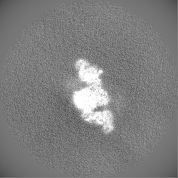
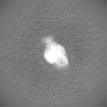
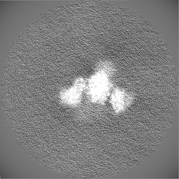
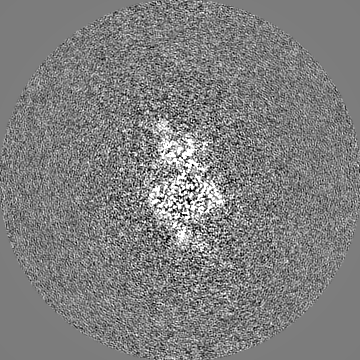
Journal: Microbiology (Reading) / Year: 2024 Title: Cryo-electron microscopy structure of the di-domain core of polyketide synthase 13, essential for mycobacterial mycolic acid synthesis. Authors: Hannah E Johnston / Sarah M Batt / Alistair K Brown / Christos G Savva / Gurdyal S Besra / Klaus Fütterer / Abstract: Mycobacteria are known for their complex cell wall, which comprises layers of peptidoglycan, polysaccharides and unusual fatty acids known as mycolic acids that form their unique outer membrane. ...Mycobacteria are known for their complex cell wall, which comprises layers of peptidoglycan, polysaccharides and unusual fatty acids known as mycolic acids that form their unique outer membrane. Polyketide synthase 13 (Pks13) of , the bacterial organism causing tuberculosis, catalyses the last step of mycolic acid synthesis prior to export to and assembly in the cell wall. Due to its essentiality, Pks13 is a target for several novel anti-tubercular inhibitors, but its 3D structure and catalytic reaction mechanism remain to be fully elucidated. Here, we report the molecular structure of the catalytic core domains of Pks13 (Mt-Pks13), determined by transmission cryo-electron microscopy (cryoEM) to a resolution of 3.4 Å. We observed a homodimeric assembly comprising the ketoacyl synthase (KS) domain at the centre, mediating dimerization, and the acyltransferase (AT) domains protruding in opposite directions from the central KS domain dimer. In addition to the KS-AT di-domains, the cryoEM map includes features not covered by the di-domain structural model that we predicted to contain a dimeric domain similar to dehydratases, yet likely lacking catalytic function. Analytical ultracentrifugation data indicate a pH-dependent equilibrium between monomeric and dimeric assembly states, while comparison with the previously determined structures of Pks13 indicates architectural flexibility. Combining the experimentally determined structure with modelling in AlphaFold2 suggests a structural scaffold with a relatively stable dimeric core, which combines with considerable conformational flexibility to facilitate the successive steps of the Claisen-type condensation reaction catalysed by Pks13.
Name: Polyketide synthase Pks13 / type: protein_or_peptide / ID: 1 / Number of copies: 2 / Enantiomer: LEVO EC number: Transferases; Acyltransferases; Transferring groups other than aminoacyl groups
pH: 7.9 / Component - Concentration: 20.0 mM / Component - Formula: C4H11NO3 / Component - Name: Tris Details: 20 mM Tris-HCl pH 7.9, 50 mM NaCl, 2.5 mM beta-mercaptoethanol
Grid
Model: Quantifoil R1.2/1.3 / Material: GOLD / Support film - Material: CARBON / Support film - topology: HOLEY / Pretreatment - Type: GLOW DISCHARGE / Pretreatment - Time: 60 sec.
Vitrification
Cryogen name: ETHANE / Chamber humidity: 100 % / Chamber temperature: 291 K / Instrument: FEI VITROBOT MARK IV
-
Electron microscopy
Microscope
FEI TITAN KRIOS
Image recording
Film or detector model: GATAN K3 (6k x 4k) / Number grids imaged: 1 / Number real images: 1412 / Average exposure time: 3.0 sec. / Average electron dose: 16.5 e/Å2 / Details: movie mode
Electron beam
Acceleration voltage: 300 kV / Electron source: FIELD EMISSION GUN
In the structure databanks used in Yorodumi, some data are registered as the other names, "COVID-19 virus" and "2019-nCoV". Here are the details of the virus and the list of structure data.
Jan 31, 2019. EMDB accession codes are about to change! (news from PDBe EMDB page)
EMDB accession codes are about to change! (news from PDBe EMDB page)
The allocation of 4 digits for EMDB accession codes will soon come to an end. Whilst these codes will remain in use, new EMDB accession codes will include an additional digit and will expand incrementally as the available range of codes is exhausted. The current 4-digit format prefixed with “EMD-” (i.e. EMD-XXXX) will advance to a 5-digit format (i.e. EMD-XXXXX), and so on. It is currently estimated that the 4-digit codes will be depleted around Spring 2019, at which point the 5-digit format will come into force.
The EM Navigator/Yorodumi systems omit the EMD- prefix.
Related info.:Q: What is EMD? / ID/Accession-code notation in Yorodumi/EM Navigator
Yorodumi is a browser for structure data from EMDB, PDB, SASBDB, etc.
This page is also the successor to EM Navigator detail page, and also detail information page/front-end page for Omokage search.
The word "yorodu" (or yorozu) is an old Japanese word meaning "ten thousand". "mi" (miru) is to see.
Related info.:EMDB / PDB / SASBDB / Comparison of 3 databanks / Yorodumi Search / Aug 31, 2016. New EM Navigator & Yorodumi / Yorodumi Papers / Jmol/JSmol / Function and homology information / Changes in new EM Navigator and Yorodumi
 Movie
Movie Controller

 Yorodumi
 Open data
 Basic information
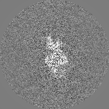
 Map data
 Sample
 Keywords
 Function and homology information
 Mycobacterium tuberculosis (bacteria) /
  Authors
 United Kingdom, 2 items
  Citation
 Structure visualization
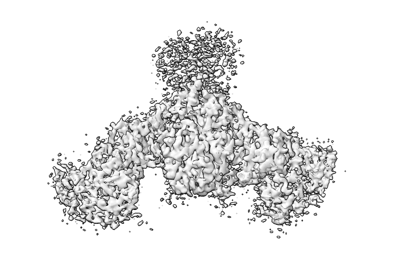
 Downloads & links
 emd_50185.png
 http://ftp.pdbj.org/pub/emdb/structures/EMD-50185


 Z (Sec.)
 Y (Row.)
 X (Col.)


 Sample components
 Processing
 Electron microscopy
 FIELD EMISSION GUN
