[English] 日本語
 Yorodumi
- PDB-9f48: KS + AT di-domain of polyketide synthase 13 in Mycobacterium tube... -
+ Open data
- Basic information
| Entry | Database: PDB / ID: 9f48 | |||||||||
|---|---|---|---|---|---|---|---|---|---|---|
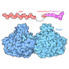
| Title | KS + AT di-domain of polyketide synthase 13 in Mycobacterium tuberculosis | |||||||||
 Components Components | Polyketide synthase Pks13 | |||||||||
 Keywords Keywords | BIOSYNTHETIC PROTEIN / Mycolic acid synthesis / Claisen condensation / cell wall synthesis | |||||||||
| Function / homology |  Function and homology information Function and homology informationDIM/DIP cell wall layer assembly / fatty acid synthase activity / phosphopantetheine binding / 3-oxoacyl-[acyl-carrier-protein] synthase activity / Transferases; Acyltransferases; Transferring groups other than aminoacyl groups / fatty acid biosynthetic process / cytoplasm Similarity search - Function | |||||||||
| Biological species |  Mycobacterium tuberculosis H37Rv (bacteria) Mycobacterium tuberculosis H37Rv (bacteria) | |||||||||
| Method | ELECTRON MICROSCOPY / single particle reconstruction / cryo EM / Resolution: 3.4 Å | |||||||||
 Authors Authors | Johnston, H.E. / Futterer, K. | |||||||||
| Funding support |  United Kingdom, 2items United Kingdom, 2items
| |||||||||
 Citation Citation |  Journal: Microbiology (Reading) / Year: 2024 Journal: Microbiology (Reading) / Year: 2024Title: Cryo-electron microscopy structure of the di-domain core of polyketide synthase 13, essential for mycobacterial mycolic acid synthesis. Authors: Hannah E Johnston / Sarah M Batt / Alistair K Brown / Christos G Savva / Gurdyal S Besra / Klaus Fütterer /  Abstract: Mycobacteria are known for their complex cell wall, which comprises layers of peptidoglycan, polysaccharides and unusual fatty acids known as mycolic acids that form their unique outer membrane. ...Mycobacteria are known for their complex cell wall, which comprises layers of peptidoglycan, polysaccharides and unusual fatty acids known as mycolic acids that form their unique outer membrane. Polyketide synthase 13 (Pks13) of , the bacterial organism causing tuberculosis, catalyses the last step of mycolic acid synthesis prior to export to and assembly in the cell wall. Due to its essentiality, Pks13 is a target for several novel anti-tubercular inhibitors, but its 3D structure and catalytic reaction mechanism remain to be fully elucidated. Here, we report the molecular structure of the catalytic core domains of Pks13 (Mt-Pks13), determined by transmission cryo-electron microscopy (cryoEM) to a resolution of 3.4 Å. We observed a homodimeric assembly comprising the ketoacyl synthase (KS) domain at the centre, mediating dimerization, and the acyltransferase (AT) domains protruding in opposite directions from the central KS domain dimer. In addition to the KS-AT di-domains, the cryoEM map includes features not covered by the di-domain structural model that we predicted to contain a dimeric domain similar to dehydratases, yet likely lacking catalytic function. Analytical ultracentrifugation data indicate a pH-dependent equilibrium between monomeric and dimeric assembly states, while comparison with the previously determined structures of Pks13 indicates architectural flexibility. Combining the experimentally determined structure with modelling in AlphaFold2 suggests a structural scaffold with a relatively stable dimeric core, which combines with considerable conformational flexibility to facilitate the successive steps of the Claisen-type condensation reaction catalysed by Pks13. | |||||||||
| History |
|
- Structure visualization
| Structure viewer | Molecule:  Molmil Molmil Jmol/JSmol Jmol/JSmol |
|---|
- Downloads & links
- Download
| PDBx/mmCIF format |  9f48.cif.gz 9f48.cif.gz | 459 KB | Display |  PDBx/mmCIF format PDBx/mmCIF format |
|---|---|---|---|---|
| PDB format |  pdb9f48.ent.gz pdb9f48.ent.gz | 287.8 KB | Display |  PDB format PDB format |
| PDBx/mmJSON format |  9f48.json.gz 9f48.json.gz | Tree view |  PDBx/mmJSON format PDBx/mmJSON format | |
| Others |  Other downloads Other downloads |
-Validation report
| Summary document |  9f48_validation.pdf.gz 9f48_validation.pdf.gz | 1.2 MB | Display |  wwPDB validaton report wwPDB validaton report |
|---|---|---|---|---|
| Full document |  9f48_full_validation.pdf.gz 9f48_full_validation.pdf.gz | 1.2 MB | Display | |
| Data in XML |  9f48_validation.xml.gz 9f48_validation.xml.gz | 58.3 KB | Display | |
| Data in CIF |  9f48_validation.cif.gz 9f48_validation.cif.gz | 87 KB | Display | |
| Arichive directory |  https://data.pdbj.org/pub/pdb/validation_reports/f4/9f48 https://data.pdbj.org/pub/pdb/validation_reports/f4/9f48 ftp://data.pdbj.org/pub/pdb/validation_reports/f4/9f48 ftp://data.pdbj.org/pub/pdb/validation_reports/f4/9f48 | HTTPS FTP |
-Related structure data
| Related structure data |  50185MC M: map data used to model this data C: citing same article ( |
|---|---|
| Similar structure data | Similarity search - Function & homology  F&H Search F&H Search |
- Links
Links
- Assembly
| Deposited unit | 
|
|---|---|
| 1 |
|
- Components
| #1: Protein | Mass: 186642.188 Da / Num. of mol.: 2 Source method: isolated from a genetically manipulated source Source: (gene. exp.)  Mycobacterium tuberculosis H37Rv (bacteria) Mycobacterium tuberculosis H37Rv (bacteria)Gene: pks13, Rv3800c / Production host:  References: UniProt: I6X8D2, Transferases; Acyltransferases; Transferring groups other than aminoacyl groups Has protein modification | N | |
|---|
-Experimental details
-Experiment
| Experiment | Method: ELECTRON MICROSCOPY |
|---|---|
| EM experiment | Aggregation state: PARTICLE / 3D reconstruction method: single particle reconstruction |
- Sample preparation
| Component | Name: Subunit of Pks13 / Type: COMPLEX / Entity ID: all / Source: RECOMBINANT |
|---|---|
| Molecular weight | Value: 0.187 MDa / Experimental value: YES |
| Source (natural) | Organism:  |
| Source (recombinant) | Organism:  |
| Buffer solution | pH: 7.9 Details: 20 mM Tris-HCl pH 7.9, 50 mM NaCl, 2.5 mM beta-mercaptoethanol |
| Buffer component | Conc.: 20 mM / Name: Tris / Formula: C4H11NO3 |
| Specimen | Conc.: 0.9 mg/ml / Embedding applied: NO / Shadowing applied: NO / Staining applied: NO / Vitrification applied: YES |
| Specimen support | Grid material: GOLD / Grid type: Quantifoil R1.2/1.3 |
| Vitrification | Instrument: FEI VITROBOT MARK IV / Cryogen name: ETHANE / Humidity: 100 % / Chamber temperature: 291 K |
- Electron microscopy imaging
| Experimental equipment |  Model: Titan Krios / Image courtesy: FEI Company |
|---|---|
| Microscopy | Model: FEI TITAN KRIOS |
| Electron gun | Electron source:  FIELD EMISSION GUN / Accelerating voltage: 300 kV / Illumination mode: FLOOD BEAM FIELD EMISSION GUN / Accelerating voltage: 300 kV / Illumination mode: FLOOD BEAM |
| Electron lens | Mode: OTHER / Nominal defocus max: 2700 nm / Nominal defocus min: 1500 nm / Cs: 2.7 mm / C2 aperture diameter: 100 µm |
| Specimen holder | Specimen holder model: FEI TITAN KRIOS AUTOGRID HOLDER |
| Image recording | Average exposure time: 3 sec. / Electron dose: 16.5 e/Å2 / Film or detector model: GATAN K3 (6k x 4k) / Num. of grids imaged: 1 / Num. of real images: 1412 / Details: movie mode |
- Processing
| EM software | Name: PHENIX / Version: 1.21.1_5286 / Category: model refinement | ||||||||||||||||||||||||
|---|---|---|---|---|---|---|---|---|---|---|---|---|---|---|---|---|---|---|---|---|---|---|---|---|---|
| CTF correction | Type: NONE | ||||||||||||||||||||||||
| Particle selection | Num. of particles selected: 310055 | ||||||||||||||||||||||||
| 3D reconstruction | Resolution: 3.4 Å / Resolution method: OTHER / Num. of particles: 168566 / Symmetry type: POINT | ||||||||||||||||||||||||
| Refinement | Cross valid method: NONE Stereochemistry target values: GeoStd + Monomer Library + CDL v1.2 | ||||||||||||||||||||||||
| Displacement parameters | Biso mean: 70.54 Å2 | ||||||||||||||||||||||||
| Refine LS restraints |
|
 Movie
Movie Controller


 PDBj
PDBj


