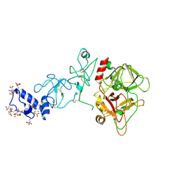
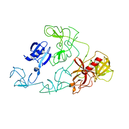
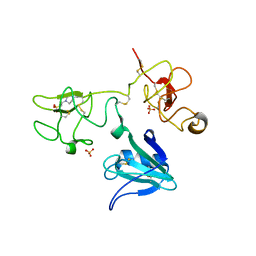
6PX5
 
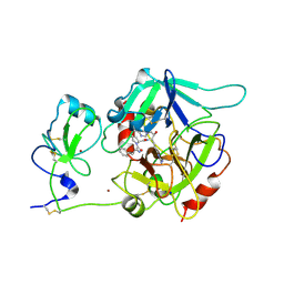 | | CRYSTAL STRUCTURE OF HUMAN MEIZOTHROMBIN DESF1 MUTANT S195A bound with PPACK | | 分子名称: | 2-acetamido-2-deoxy-beta-D-glucopyranose-(1-4)-2-acetamido-2-deoxy-beta-D-glucopyranose, D-phenylalanyl-N-[(2S,3S)-6-{[amino(iminio)methyl]amino}-1-chloro-2-hydroxyhexan-3-yl]-L-prolinamide, Prothrombin, ... | | 著者 | Pelc, L.A, Koester, S.K, Chen, Z, Gistover, N, Di Cera, E. | | 登録日 | 2019-07-24 | | 公開日 | 2019-09-04 | | 最終更新日 | 2023-10-11 | | 実験手法 | X-RAY DIFFRACTION (2.4 Å) | | 主引用文献 | Residues W215, E217 and E192 control the allosteric E*-E equilibrium of thrombin.
Sci Rep, 9, 2019
|
|
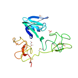
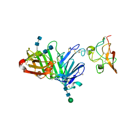
1NL2
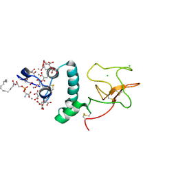 | | BOVINE PROTHROMBIN FRAGMENT 1 IN COMPLEX WITH CALCIUM AND LYSOPHOSPHOTIDYLSERINE | | 分子名称: | 2-acetamido-2-deoxy-beta-D-glucopyranose, 2-acetamido-2-deoxy-beta-D-glucopyranose-(1-4)-2-acetamido-2-deoxy-beta-D-glucopyranose, CALCIUM ION, ... | | 著者 | Huang, M, Huang, G, Furie, B, Seaton, B, Furie, B.C. | | 登録日 | 2003-01-06 | | 公開日 | 2003-09-16 | | 最終更新日 | 2023-11-15 | | 実験手法 | X-RAY DIFFRACTION (2.3 Å) | | 主引用文献 | Structural basis of membrane binding by Gla domains of vitamin K-dependent proteins.
Nat.Struct.Biol., 10, 2003
|
|
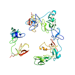
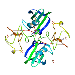
2DOI
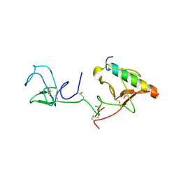 | | The X-ray crystallographic structure of the angiogenesis inhibitor, angiostatin, bound to a peptide from the group A streptococcus protein PAM | | 分子名称: | Angiostatin, Plasminogen-binding group A streptococcal M-like protein PAM | | 著者 | Cnudde, S.E, Prorok, M, Castellino, F.J, Geiger, J.H. | | 登録日 | 2006-04-29 | | 公開日 | 2006-12-05 | | 最終更新日 | 2023-10-25 | | 実験手法 | X-RAY DIFFRACTION (3.1 Å) | | 主引用文献 | X-ray crystallographic structure of the angiogenesis inhibitor, angiostatin, bound to a peptide from the group A streptococcal surface protein PAM
Biochemistry, 45, 2006
|
|
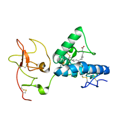
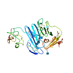
2DOH
 | | The X-ray crystallographic structure of the angiogenesis inhibitor, angiostatin, bound a to a peptide from the group A streptococcal surface protein PAM | | 分子名称: | 1,4-DIETHYLENE DIOXIDE, Angiostatin, Plasminogen-binding group A streptococcal M-like protein PAM | | 著者 | Cnudde, S.E, Prorok, M, Castellino, F.J, Geiger, J.H. | | 登録日 | 2006-04-29 | | 公開日 | 2006-12-05 | | 最終更新日 | 2023-10-25 | | 実験手法 | X-RAY DIFFRACTION (2.3 Å) | | 主引用文献 | X-ray crystallographic structure of the angiogenesis inhibitor, angiostatin, bound to a peptide from the group A streptococcal surface protein PAM
Biochemistry, 45, 2006
|
|
7ME4
 | | Structure of the extracellular WNT-binding module in Drosophila Ror2/Nrk | | 分子名称: | 2-acetamido-2-deoxy-beta-D-glucopyranose-(1-4)-2-acetamido-2-deoxy-beta-D-glucopyranose, PALMITOLEIC ACID, Tyrosine-protein kinase transmembrane receptor Ror2 | | 著者 | Mendrola, J.M, Shi, F, Perry, K, Stayrook, S.E, Lemmon, M.A. | | 登録日 | 2021-04-06 | | 公開日 | 2021-10-13 | | 最終更新日 | 2023-10-18 | | 実験手法 | X-RAY DIFFRACTION (1.75 Å) | | 主引用文献 | ROR and RYK extracellular region structures suggest that receptor tyrosine kinases have distinct WNT-recognition modes.
Cell Rep, 37, 2021
|
|
1I5K
 | | STRUCTURE AND BINDING DETERMINANTS OF THE RECOMBINANT KRINGLE-2 DOMAIN OF HUMAN PLASMINOGEN TO AN INTERNAL PEPTIDE FROM A GROUP A STREPTOCOCCAL SURFACE PROTEIN | | 分子名称: | M PROTEIN, PLASMINOGEN | | 著者 | Rios-Steiner, J.L, Schenone, M, Mochalkin, I, Tulinsky, A, Castellino, F.J. | | 登録日 | 2001-02-27 | | 公開日 | 2001-08-01 | | 最終更新日 | 2023-08-09 | | 実験手法 | X-RAY DIFFRACTION (2.7 Å) | | 主引用文献 | Structure and binding determinants of the recombinant kringle-2 domain of human plasminogen to an internal peptide from a group A Streptococcal surface protein.
J.Mol.Biol., 308, 2001
|
|
2HPP
 | |
2HPQ
 | |
5EDM
 | | Crystal structure of prothrombin deletion mutant residues 154-167 ( Form I ) | | 分子名称: | 2-acetamido-2-deoxy-beta-D-glucopyranose, 2-acetamido-2-deoxy-beta-D-glucopyranose-(1-4)-2-acetamido-2-deoxy-beta-D-glucopyranose, GLYCEROL, ... | | 著者 | Pozzi, N, Chen, Z, Di Cera, E. | | 登録日 | 2015-10-21 | | 公開日 | 2016-01-20 | | 最終更新日 | 2023-11-15 | | 実験手法 | X-RAY DIFFRACTION (2.2 Å) | | 主引用文献 | How the Linker Connecting the Two Kringles Influences Activation and Conformational Plasticity of Prothrombin.
J.Biol.Chem., 291, 2016
|
|
5EDK
 | | Crystal structure of prothrombin deletion mutant residues 146-167 ( Form II ). | | 分子名称: | 2-acetamido-2-deoxy-beta-D-glucopyranose, 2-acetamido-2-deoxy-beta-D-glucopyranose-(1-4)-2-acetamido-2-deoxy-beta-D-glucopyranose, MAGNESIUM ION, ... | | 著者 | Pozzi, N, Chen, Z, Di Cera, E. | | 登録日 | 2015-10-21 | | 公開日 | 2016-01-20 | | 最終更新日 | 2023-11-15 | | 実験手法 | X-RAY DIFFRACTION (3.214 Å) | | 主引用文献 | How the Linker Connecting the Two Kringles Influences Activation and Conformational Plasticity of Prothrombin.
J.Biol.Chem., 291, 2016
|
|
3HN4
 | |
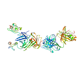
8OS5
 | | Crystal structure of the Factor XII heavy chain reveals an interlocking dimer with a FnII to kringle domain interaction | | 分子名称: | Coagulation factor XII-Mie | | 著者 | Li, C, Saleem, M, Kaira, B.G, Brown, A, Wilson, C, Philippou, H, Emsley, J. | | 登録日 | 2023-04-18 | | 公開日 | 2024-03-27 | | 実験手法 | X-RAY DIFFRACTION (3.4 Å) | | 主引用文献 | Factor XII and kininogen asymmetric assembly with gC1qR/C1QBP/P32 is governed by allostery.
Blood, 136, 2020
|
|
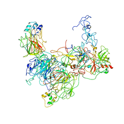
9CTH
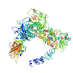 | | Preliminary map of the Prothrombin-prothrombinase complex on nano discs | | 分子名称: | 2-acetamido-2-deoxy-beta-D-glucopyranose, Activated Factor V (FVa) heavy chain, Activated Factor V (FVa) light chain, ... | | 著者 | Stojanovski, B.M, Mohammed, B.M, Di Cera, E. | | 登録日 | 2024-07-25 | | 公開日 | 2024-08-07 | | 最終更新日 | 2024-08-14 | | 実験手法 | ELECTRON MICROSCOPY (6.47 Å) | | 主引用文献 | The Prothrombin-Prothrombinase Interaction.
Subcell Biochem, 104, 2024
|
|
4D3C
 | |
7TPP
 | |
3SP8
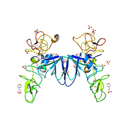 | | Crystal structure of NK2 in complex with fractionated Heparin DP10 | | 分子名称: | (4R)-2-METHYLPENTANE-2,4-DIOL, (4S)-2-METHYL-2,4-PENTANEDIOL, 2-(N-MORPHOLINO)-ETHANESULFONIC ACID, ... | | 著者 | Recacha, R, Mulloy, B, Gherardi, E. | | 登録日 | 2011-07-01 | | 公開日 | 2012-07-04 | | 最終更新日 | 2023-09-13 | | 実験手法 | X-RAY DIFFRACTION (1.86 Å) | | 主引用文献 | Crystal structure of NK2 in complex with fractionated Heparin DP10
TO BE PUBLISHED
|
|
4DUR
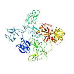 | | The X-ray Crystal Structure of Full-Length type II Human Plasminogen | | 分子名称: | ACETATE ION, BICARBONATE ION, CHLORIDE ION, ... | | 著者 | Law, R.H.P, Caradoc-Davies, T, Whisstock, J.C. | | 登録日 | 2012-02-22 | | 公開日 | 2012-03-28 | | 最終更新日 | 2023-11-08 | | 実験手法 | X-RAY DIFFRACTION (2.45 Å) | | 主引用文献 | The X-ray crystal structure of full-length human plasminogen
Cell Rep, 1, 2012
|
|
4DUU
 | |
3U73
 | | Crystal structure of stabilized human uPAR mutant in complex with ATF | | 分子名称: | 2-acetamido-2-deoxy-beta-D-glucopyranose-(1-4)-2-acetamido-2-deoxy-beta-D-glucopyranose, Urokinase plasminogen activator surface receptor, Urokinase-type plasminogen activator, ... | | 著者 | Huang, M.D, Xu, X, Yuan, C. | | 登録日 | 2011-10-13 | | 公開日 | 2012-04-18 | | 最終更新日 | 2020-07-29 | | 実験手法 | X-RAY DIFFRACTION (3.19 Å) | | 主引用文献 | Crystal structure of the urokinase receptor in a ligand-free form.
J.Mol.Biol., 416, 2012
|
|
3BT2
 | | Structure of urokinase receptor, urokinase and vitronectin complex | | 分子名称: | 2-acetamido-2-deoxy-beta-D-glucopyranose, 2-acetamido-2-deoxy-beta-D-glucopyranose-(1-4)-2-acetamido-2-deoxy-beta-D-glucopyranose, Urokinase plasminogen activator surface receptor, ... | | 著者 | Huang, M. | | 登録日 | 2007-12-27 | | 公開日 | 2008-03-25 | | 最終更新日 | 2023-11-01 | | 実験手法 | X-RAY DIFFRACTION (2.5 Å) | | 主引用文献 | Crystal structures of two human vitronectin, urokinase and urokinase receptor complexes
Nat.Struct.Mol.Biol., 15, 2008
|
|
3BT1
 | | Structure of urokinase receptor, urokinase and vitronectin complex | | 分子名称: | 2-acetamido-2-deoxy-beta-D-glucopyranose, Urokinase plasminogen activator surface receptor, Urokinase-type plasminogen activator, ... | | 著者 | Huang, M. | | 登録日 | 2007-12-27 | | 公開日 | 2008-03-25 | | 最終更新日 | 2023-11-01 | | 実験手法 | X-RAY DIFFRACTION (2.8 Å) | | 主引用文献 | Crystal structures of two human vitronectin, urokinase and urokinase receptor complexes
Nat.Struct.Mol.Biol., 15, 2008
|
|
1BHT
 | | NK1 FRAGMENT OF HUMAN HEPATOCYTE GROWTH FACTOR | | 分子名称: | 4-(2-HYDROXYETHYL)-1-PIPERAZINE ETHANESULFONIC ACID, HEPATOCYTE GROWTH FACTOR, SULFATE ION | | 著者 | Ultsch, M.H, Lokker, N.A, Godowski, P.J, De Vos, A.M. | | 登録日 | 1998-06-10 | | 公開日 | 1998-11-04 | | 最終更新日 | 2011-07-13 | | 実験手法 | X-RAY DIFFRACTION (2 Å) | | 主引用文献 | Crystal structure of the NK1 fragment of human hepatocyte growth factor at 2.0 A resolution.
Structure, 6, 1998
|
|
3LAQ
 | | Structure-based engineering of species selectivity in the uPA-uPAR interaction | | 分子名称: | 2-acetamido-2-deoxy-beta-D-glucopyranose, Urokinase plasminogen activator surface receptor, Urokinase-type plasminogen activator | | 著者 | Huang, M. | | 登録日 | 2010-01-06 | | 公開日 | 2010-02-02 | | 最終更新日 | 2020-07-29 | | 実験手法 | X-RAY DIFFRACTION (3.2 Å) | | 主引用文献 | Structure-based engineering of species selectivity in the interaction between urokinase and its receptor: implication for preclinical cancer therapy.
J.Biol.Chem., 285, 2010
|
|
4IUA
 | | Crystal Structure of the NK2 Fragment (31-290) of the mouse Hepatocyte Growth Factor/Scatter Factor | | 分子名称: | 4-(2-HYDROXYETHYL)-1-PIPERAZINE ETHANESULFONIC ACID, Hepatocyte growth factor, SULFATE ION | | 著者 | Tolbert, W.D, Zhou, E, Kovach, A, Melcher, K, Xu, H.E. | | 登録日 | 2013-01-20 | | 公開日 | 2013-02-13 | | 最終更新日 | 2023-09-20 | | 実験手法 | X-RAY DIFFRACTION (3.05 Å) | | 主引用文献 | Crystal Structure of the NK2 Fragment of the mouse Hepatocyte Growth Factor/Scatter Factor
To be Published
|
|
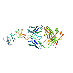
4K24
 | | Structure of anti-uPAR Fab ATN-658 in complex with uPAR | | 分子名称: | 2-acetamido-2-deoxy-beta-D-glucopyranose, 2-acetamido-2-deoxy-beta-D-glucopyranose-(1-4)-2-acetamido-2-deoxy-beta-D-glucopyranose, Urokinase plasminogen activator surface receptor, ... | | 著者 | Huang, M.D, Xu, X, Yuan, C. | | 登録日 | 2013-04-08 | | 公開日 | 2014-02-26 | | 最終更新日 | 2023-11-08 | | 実験手法 | X-RAY DIFFRACTION (4.5 Å) | | 主引用文献 | Identification of a New Epitope in uPAR as a Target for the Cancer Therapeutic Monoclonal Antibody ATN-658, a Structural Homolog of the uPAR Binding Integrin CD11b ( alpha M)
Plos One, 9, 2014
|
|


