Loading
PDBj
MenuPDBj@FacebookPDBj@TwitterPDBj@YouTubewwPDB FoundationwwPDB
RCSB PDBPDBeBMRBAdv. SearchSearch help
PDB: 274 results

6XVX
DownloadVisualize
BU of 6xvx by Molmil
X-ray structure obtained upon reaction of dirhodium tetraacetate with RNase A (high resolution)
Descriptor: ACETATE ION, RHODIUM(III) ION, Ribonuclease pancreatic
Authors:Merlino, A, Ferraro, G.
Deposit date:2020-01-22
Release date:2020-02-19
Last modified:2024-01-24
Method:X-RAY DIFFRACTION (1.4 Å)
Cite:Protein interactions of dirhodium tetraacetate: a structural study.
Dalton Trans, 49, 2020
6XW0
DownloadVisualize
BU of 6xw0 by Molmil
X-ray structure obtained upon reaction of dirhodium tetraacetate with RNase A (low resolution)
Descriptor: ACETATE ION, RHODIUM(III) ION, Ribonuclease pancreatic
Authors:Merlino, A, Ferraro, G.
Deposit date:2020-01-22
Release date:2020-02-19
Last modified:2024-01-24
Method:X-RAY DIFFRACTION (1.8 Å)
Cite:Protein interactions of dirhodium tetraacetate: a structural study.
Dalton Trans, 49, 2020
3LJF
DownloadVisualize
BU of 3ljf by Molmil
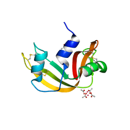
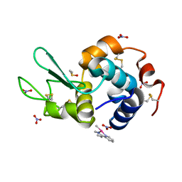
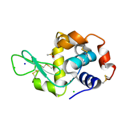
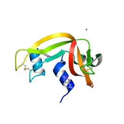
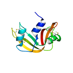
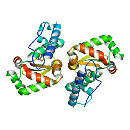
The X-ray structure of iron superoxide dismutase from Pseudoalteromonas haloplanktis (crystal form II)
Descriptor: FE (III) ION, alpha-D-glucopyranose-(1-1)-alpha-D-glucopyranose, iron superoxide dismutase
Authors:Merlino, A, Russo Krauss, I, Rossi, B, Conte, M, Vergara, A, Sica, F.
Deposit date:2010-01-26
Release date:2010-09-08
Last modified:2023-09-06
Method:X-RAY DIFFRACTION (2.1 Å)
Cite:Structure and flexibility in cold-adapted iron superoxide dismutases: the case of the enzyme isolated from Pseudoalteromonas haloplanktis.
J.Struct.Biol., 172, 2010
5ILC
DownloadVisualize
BU of 5ilc by Molmil
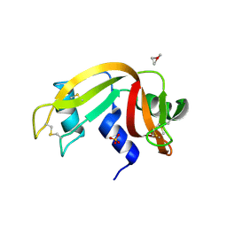
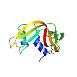
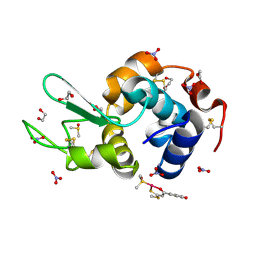
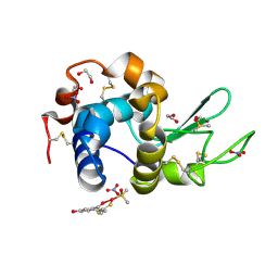
The X-ray structure of the adduct formed in the reaction between hen egg white lysozyme a compound 2, a platin(II) compound containing a O, S bidentate ligand
Descriptor: ACETATE ION, CHLORIDE ION, DIMETHYL SULFOXIDE, ...
Authors:Merlino, A, Ferraro, G.
Deposit date:2016-03-04
Release date:2016-12-07
Last modified:2024-01-10
Method:X-RAY DIFFRACTION (1.75 Å)
Cite:Platinum(ii) O,S complexes as potential metallodrugs against Cisplatin resistance.
Dalton Trans, 45, 2016
6RJV
DownloadVisualize
BU of 6rjv by Molmil
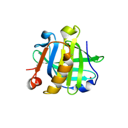
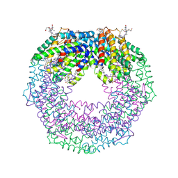
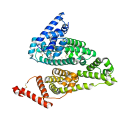
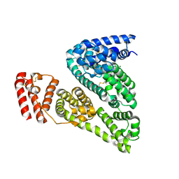
The X-ray structure of the Gold/Serum Albumin adduct obtained upon reaction of the protein with AuL12, a gold(III) dithiocarbamate complex
Descriptor: GOLD ION, MAGNESIUM ION, Serum albumin
Authors:Merlino, A, Giorgio, A, Ferraro, G.
Deposit date:2019-04-29
Release date:2019-08-14
Last modified:2024-01-24
Method:X-RAY DIFFRACTION (3.21 Å)
Cite:Structural Characterization of a Gold/Serum Albumin Complex.
Inorg.Chem., 58, 2019
6QEA
DownloadVisualize
BU of 6qea by Molmil
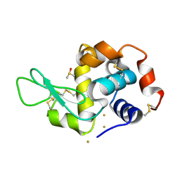
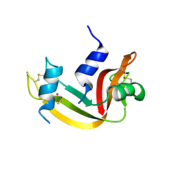
The X-ray structure of the adduct formed in the reaction between hen egg white lysozyme and complex I, a pentacoordinate Pt(II) compound containing 2,9-dimethyl-1,10-phenanthroline, dimethylfumarate, methyl and iodine as ligands
Descriptor: DIMETHYL SULFOXIDE, Lysozyme C, NITRATE ION, ...
Authors:Merlino, A, Ferraro, G.
Deposit date:2019-01-07
Release date:2019-03-27
Last modified:2024-01-24
Method:X-RAY DIFFRACTION (1.96 Å)
Cite:Reaction with Proteins of a Five-Coordinate Platinum(II) Compound.
Int J Mol Sci, 20, 2019
6QE9
DownloadVisualize
BU of 6qe9 by Molmil
The X-ray structure of the adduct formed in the reaction between bovine pancreatic ribonuclease and complex I, a pentacoordinate Pt(II) compound containing 2,9-dimethyl-1,10-phenanthroline, dimethylfumarate, methyl and iodine as ligands
Descriptor: Ribonuclease pancreatic, SULFATE ION, pentacoordinate Pt(II) compound
Authors:Merlino, A, Ferraro, G.
Deposit date:2019-01-07
Release date:2019-02-27
Last modified:2024-01-24
Method:X-RAY DIFFRACTION (2.03 Å)
Cite:Reaction with Proteins of a Five-Coordinate Platinum(II) Compound.
Int J Mol Sci, 20, 2019
7OR6
DownloadVisualize
BU of 7or6 by Molmil
The crystal structure of the domain-swapped dimer of onconase
Descriptor: Protein P-30
Authors:Merlino, A, Loreto, D.
Deposit date:2021-06-04
Release date:2021-10-06
Last modified:2024-01-31
Method:X-RAY DIFFRACTION (2.12 Å)
Cite:The crystal structure of the domain-swapped dimer of onconase highlights some catalytic and antitumor activity features of the enzyme.
Int.J.Biol.Macromol., 191, 2021
7ORD
DownloadVisualize
BU of 7ord by Molmil
The crystal structure of the domain-swapped dimer of onconase (2)
Descriptor: ACETATE ION, Protein P-30, SULFATE ION
Authors:Merlino, A, Loreto, D.
Deposit date:2021-06-05
Release date:2021-10-06
Last modified:2024-01-31
Method:X-RAY DIFFRACTION (2.14 Å)
Cite:The crystal structure of the domain-swapped dimer of onconase highlights some catalytic and antitumor activity features of the enzyme.
Int.J.Biol.Macromol., 191, 2021
7NQB
DownloadVisualize
BU of 7nqb by Molmil
X.ray structure of the oxaliplatin/beta-lactoglobulin adduct
Descriptor: Beta-lactoglobulin, PLATINUM (II) ION
Authors:Merlino, A, Loreto, D, Ferraro, G.
Deposit date:2021-03-01
Release date:2022-03-23
Last modified:2024-01-31
Method:X-RAY DIFFRACTION (2.01 Å)
Cite:Protein-Based Delivery Systems for Anticancer Metallodrugs: Structure and Biological Activity of the Oxaliplatin/ beta-Lactoglobulin Adduct.
Pharmaceuticals, 15, 2022
7R1Q
DownloadVisualize
BU of 7r1q by Molmil
X-ray structure of the adduct formed upon reaction of the gold(I) N-heterocyclic carbene complex Au2 with lysozyme
Descriptor: DIMETHYL SULFOXIDE, GOLD ION, Lysozyme
Authors:Merlino, A, Ferraro, G.
Deposit date:2022-02-03
Release date:2022-07-13
Last modified:2024-01-31
Method:X-RAY DIFFRACTION (1.1 Å)
Cite:Halo complexes of gold(I) containing glycoconjugate carbene ligands: synthesis, characterization, cytotoxicity and interaction with proteins and DNA model systems.
Dalton Trans, 51, 2022
7R1P
DownloadVisualize
BU of 7r1p by Molmil
X-ray structure of the adduct formed upon reaction of the gold(I) N-heterocyclic carbene complex Au1 with RNase A
Descriptor: GOLD ION, Ribonuclease pancreatic, SULFATE ION
Authors:Merlino, A, Ferraro, G.
Deposit date:2022-02-03
Release date:2022-07-13
Last modified:2024-01-31
Method:X-RAY DIFFRACTION (1.42 Å)
Cite:Halo complexes of gold(I) containing glycoconjugate carbene ligands: synthesis, characterization, cytotoxicity and interaction with proteins and DNA model systems.
Dalton Trans, 51, 2022
5ILF
DownloadVisualize
BU of 5ilf by Molmil
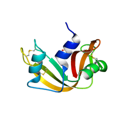
The X-ray structure of the adduct formed in the reaction between hen egg white lysozyme and compound 4, a platin(II) compound containing a O, S bidentate ligand
Descriptor: ACETATE ION, CHLORIDE ION, DIMETHYL SULFOXIDE, ...
Authors:Merlino, A, Ferraro, G.
Deposit date:2016-03-04
Release date:2016-12-07
Last modified:2024-01-10
Method:X-RAY DIFFRACTION (1.85 Å)
Cite:Platinum(ii) O,S complexes as potential metallodrugs against Cisplatin resistance.
Dalton Trans, 45, 2016
7QWH
DownloadVisualize
BU of 7qwh by Molmil
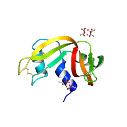
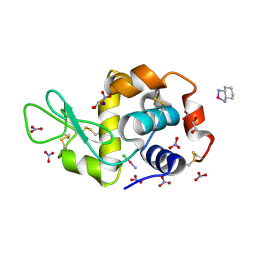
X-ray structure of the adduct formed upon reaction of a vanadium hydroxyquinoline complex with RNase A
Descriptor: 2,2-bis($l^{1}-oxidanyl)-3-oxa-1$l^{4}-aza-2$l^{4}-vanadatricyclo[6.3.1.0^{4,12}]dodeca-1(12),4,6,8,10-pentaene, Ribonuclease pancreatic
Authors:Merlino, A, Ferraro, G.
Deposit date:2022-01-25
Release date:2023-02-01
Last modified:2024-02-07
Method:X-RAY DIFFRACTION (1.573 Å)
Cite:Interaction of VIVO-8-hydroxyquinoline species with RNase A: the effect of metal ligands in the protein adduct stabilization
Inorg Chem Front, 2023
8B4N
DownloadVisualize
BU of 8b4n by Molmil
X-ray structure of phycoerythrin from Porphyridium cruentum
Descriptor: B-phycoerythrin alpha chain, B-phycoerythrin beta chain, N-METHYL ASPARAGINE, ...
Authors:Merlino, A, Ferraro, G.
Deposit date:2022-09-20
Release date:2023-02-08
Last modified:2024-02-07
Method:X-RAY DIFFRACTION (1.6 Å)
Cite:Inside out Porphyridium cruentum : Beyond the Conventional Biorefinery Concept.
Acs Sustain Chem Eng, 11, 2023
4S0Q
DownloadVisualize
BU of 4s0q by Molmil
The X-ray structure of the adduct formed in the reaction between bovine pancreatic ribonuclease and carboplatin
Descriptor: Ribonuclease pancreatic, carboplatin
Authors:Merlino, A.
Deposit date:2015-01-04
Release date:2015-11-18
Last modified:2023-09-20
Method:X-RAY DIFFRACTION (2.09 Å)
Cite:Interactions of carboplatin and oxaliplatin with proteins: Insights from X-ray structures and mass spectrometry studies of their ribonuclease A adducts.
J.Inorg.Biochem., 153, 2015
4S18
DownloadVisualize
BU of 4s18 by Molmil
The X-ray structure of the adduct formed in the reaction between bovine pancreatic ribonuclease and oxaliplatin
Descriptor: CYCLOHEXANE-1(R),2(R)-DIAMINE-PLATINUM(II), Ribonuclease pancreatic
Authors:Merlino, A.
Deposit date:2015-01-09
Release date:2015-11-25
Last modified:2023-09-20
Method:X-RAY DIFFRACTION (2.27 Å)
Cite:Interactions of carboplatin and oxaliplatin with proteins: Insights from X-ray structures and mass spectrometry studies of their ribonuclease A adducts.
J.Inorg.Biochem., 153, 2015
4Z41
DownloadVisualize
BU of 4z41 by Molmil
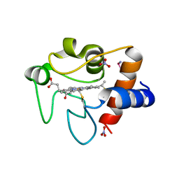
X-ray structure of the adduct formed in the reaction between lysozyme and a platinum(II) Compound with a S,O Bidentate Ligand (9a=Chloro-(1-(3'-hydroxy)-3-(methylthio)-3-thioxo-prop-1-en-1-olate-O,S)-(dimethylsulfoxide-S)-platinum(II))
Descriptor: 1,2-ETHANEDIOL, 3-[2-chloranyl-2-[dimethyl(oxidanyl)-{4}-sulfanyl]-4-ethylsulfanyl-1-oxa-3{3}-thia-2{4}-platinacyclohexa-3,5-dien-6-yl]phenol, DIMETHYL SULFOXIDE, ...
Authors:Merlino, A.
Deposit date:2015-04-01
Release date:2015-09-02
Last modified:2024-01-10
Method:X-RAY DIFFRACTION (1.89 Å)
Cite:Platinum(II) Complexes with O,S Bidentate Ligands: Biophysical Characterization, Antiproliferative Activity, and Crystallographic Evidence of Protein Binding.
Inorg.Chem., 54, 2015
4RSZ
DownloadVisualize
BU of 4rsz by Molmil
The X-ray structure of the Primary Adduct formed in the Reaction between Cisplatin and Cytochrome c
Descriptor: Cisplatin, Cytochrome c, HEME C, ...
Authors:Merlino, A.
Deposit date:2014-11-12
Release date:2015-01-14
Last modified:2023-09-20
Method:X-RAY DIFFRACTION (2.19 Å)
Cite:The X-ray structure of the primary adducts formed in the reaction between cisplatin and cytochrome c.
Chem.Commun.(Camb.), 51, 2015
4S1Y
DownloadVisualize
BU of 4s1y by Molmil
X-ray structure of human serum albumin complexed with cisplatin
Descriptor: Cisplatin, Serum albumin
Authors:Merlino, A.
Deposit date:2015-01-16
Release date:2015-04-29
Last modified:2023-09-20
Method:X-RAY DIFFRACTION (3.16 Å)
Cite:Cisplatin binding to human serum albumin: a structural study.
Chem.Commun.(Camb.), 51, 2015
5E5E
DownloadVisualize
BU of 5e5e by Molmil
X-ray structure of the adduct formed in the reaction between RNase A and a neutral organometallic derivative [Pt(pbi)(Me)(DMSO)], pbi=2-(2'-pyridil)benzimidazole (compound 3)
Descriptor: DIMETHYL SULFOXIDE, PLATINUM (II) ION, Ribonuclease pancreatic
Authors:Merlino, A.
Deposit date:2015-10-08
Release date:2015-12-09
Last modified:2016-01-13
Method:X-RAY DIFFRACTION (1.98 Å)
Cite:Cytotoxic properties of a new organometallic platinum(ii) complex and its gold(i) heterobimetallic derivatives.
Dalton Trans, 45, 2015
4Z3M
DownloadVisualize
BU of 4z3m by Molmil
X-ray structure of the adduct formed in the reaction between lysozyme and a platinum(II) Complex with S,O Bidentate Ligands (9b)
Descriptor: 1,2-ETHANEDIOL, 3-[2-chloranyl-2-[dimethyl(oxidanyl)-{4}-sulfanyl]-4-ethylsulfanyl-1-oxa-3{3}-thia-2{4}-platinacyclohexa-3,5-dien-6-yl]phenol, DIMETHYL SULFOXIDE, ...
Authors:Merlino, A.
Deposit date:2015-03-31
Release date:2015-09-02
Last modified:2024-01-10
Method:X-RAY DIFFRACTION (2.15 Å)
Cite:Platinum(II) Complexes with O,S Bidentate Ligands: Biophysical Characterization, Antiproliferative Activity, and Crystallographic Evidence of Protein Binding.
Inorg.Chem., 54, 2015
5E5F
DownloadVisualize
BU of 5e5f by Molmil
X-ray structure of the complex between RNase A and compound 4-PF6 ([(PPh3)Au(mi-pbi)Pt(Me)(DMSO)][PF6]), the heterobimetallic derivative obtained in the reaction between the organometallic compound [Pt(pbi)(Me)(DMSO)], pbi=2-(2'-pyridil)benzimidazole (compound 3) and the gold(I) compound [Au(Ph3P)][PF6]
Descriptor: ACETATE ION, GOLD ION, Ribonuclease pancreatic, ...
Authors:Merlino, A.
Deposit date:2015-10-08
Release date:2015-12-09
Last modified:2019-11-20
Method:X-RAY DIFFRACTION (1.68 Å)
Cite:Cytotoxic properties of a new organometallic platinum(ii) complex and its gold(i) heterobimetallic derivatives.
Dalton Trans, 45, 2015
4Z46
DownloadVisualize
BU of 4z46 by Molmil
X-ray structure of the bis-platinum lysozyme adduct formed in the reaction between the protein and the two drugs Cisplatin and Oxaliplatin
Descriptor: 1,2-ETHANEDIOL, CYCLOHEXANE-1(R),2(R)-DIAMINE-PLATINUM(II), Cisplatin, ...
Authors:Merlino, A.
Deposit date:2015-04-01
Release date:2015-05-27
Last modified:2015-06-10
Method:X-RAY DIFFRACTION (1.85 Å)
Cite:Oxaliplatin vs. cisplatin: competition experiments on their binding to lysozyme.
Dalton Trans, 44, 2015
3LJ9
DownloadVisualize
BU of 3lj9 by Molmil
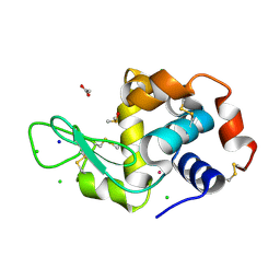
X-ray structure of the iron superoxide dismutase from pseudoalteromonas haloplanktis in complex with sodium azide
Descriptor: AZIDE ION, FE (III) ION, alpha-D-glucopyranose-(1-1)-alpha-D-glucopyranose, ...
Authors:Merlino, A, Russo Krauss, I, Rossi, B, Conte, M, Vergara, A, Sica, F.
Deposit date:2010-01-26
Release date:2010-09-08
Last modified:2023-09-06
Method:X-RAY DIFFRACTION (2.1 Å)
Cite:Structure and flexibility in cold-adapted iron superoxide dismutases: the case of the enzyme isolated from Pseudoalteromonas haloplanktis.
J.Struct.Biol., 172, 2010

222624

PDB entries from 2024-07-17

PDB statisticsPDBj update infoContact PDBjnumon