5AYS
 
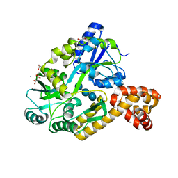
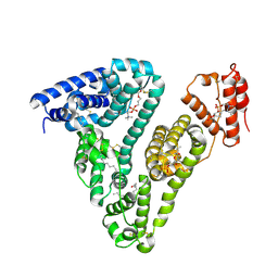
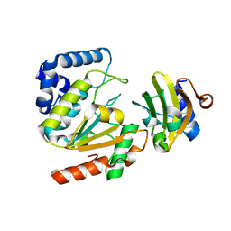 | | Crystal structure of SAUGI/HSV UDG complex | | 分子名称: | Uncharacterized protein, Uracil-DNA glycosylase | | 著者 | Wang, H.C, Ko, T.P, Huang, M.F, Wang, A.H.J. | | 登録日 | 2015-09-02 | | 公開日 | 2016-06-08 | | 最終更新日 | 2023-11-08 | | 実験手法 | X-RAY DIFFRACTION (2.09 Å) | | 主引用文献 | Using structural-based protein engineering to modulate the differential inhibition effects of SAUGI on human and HSV uracil DNA glycosylase.
Nucleic Acids Res., 44, 2016
|
|
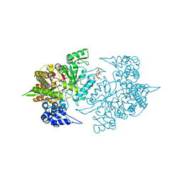
5EW6
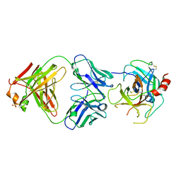
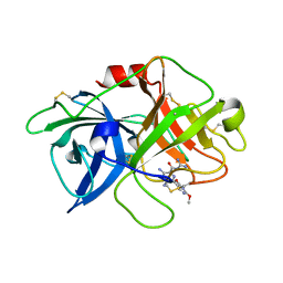
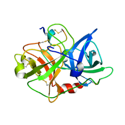
 | | Structure of ligand binding region of uPARAP at pH 7.4 without calcium | | 分子名称: | 2-acetamido-2-deoxy-beta-D-glucopyranose, 2-acetamido-2-deoxy-beta-D-glucopyranose-(1-4)-2-acetamido-2-deoxy-beta-D-glucopyranose-(1-4)-2-acetamido-2-deoxy-beta-D-glucopyranose, C-type mannose receptor 2, ... | | 著者 | Yuan, C, Huang, M. | | 登録日 | 2015-11-20 | | 公開日 | 2016-08-10 | | 最終更新日 | 2023-11-08 | | 実験手法 | X-RAY DIFFRACTION (2.29 Å) | | 主引用文献 | Crystal structures of the ligand-binding region of uPARAP: effect of calcium ion binding
Biochem.J., 473, 2016
|
|
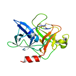
4DW2
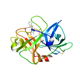
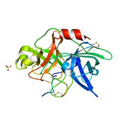
 | | The crystal structure of uPA in complex with the Fab fragment of mAb-112 | | 分子名称: | Fab fragment of pro-uPA antibody mAb-112, SULFATE ION, Urokinase-type plasminogen activator | | 著者 | Jiang, L, Botkjaer, K.A, Andersen, L.M, Yuan, C, Andreasen, P.A, Huang, M. | | 登録日 | 2012-02-24 | | 公開日 | 2013-01-16 | | 実験手法 | X-RAY DIFFRACTION (2.97 Å) | | 主引用文献 | Rezymogenation of active urokinase induced by an inhibitory antibody.
Biochem.J., 449, 2013
|
|
5E4K
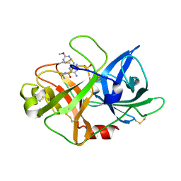
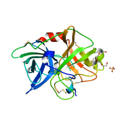
 | | Structure of ligand binding region of uPARAP at pH 7.4 | | 分子名称: | 2-acetamido-2-deoxy-beta-D-glucopyranose, 3,6,9,12,15,18,21,24-OCTAOXAHEXACOSAN-1-OL, C-type mannose receptor 2, ... | | 著者 | Yuan, C, Huang, M. | | 登録日 | 2015-10-06 | | 公開日 | 2016-08-10 | | 最終更新日 | 2023-11-08 | | 実験手法 | X-RAY DIFFRACTION (2.58 Å) | | 主引用文献 | Crystal structures of the ligand-binding region of uPARAP: effect of calcium ion binding
Biochem.J., 473, 2016
|
|
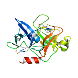
4DVA
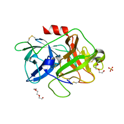 | | The crystal structure of human urokinase-type plasminogen activator catalytic domain | | 分子名称: | HEXAETHYLENE GLYCOL, SULFATE ION, Urokinase-type plasminogen activator | | 著者 | Jiang, L, Botkjaer, K.A, Andersen, L.M, Yuan, C, Andreasen, P.A, Huang, M. | | 登録日 | 2012-02-23 | | 公開日 | 2013-01-16 | | 実験手法 | X-RAY DIFFRACTION (1.94 Å) | | 主引用文献 | Rezymogenation of active urokinase induced by an inhibitory antibody
Biochem.J., 449, 2013
|
|
4ZKR
 | |
4ZKS
 | |
4ZKN
 | |
6A8G
 | | The crystal structure of muPAin-1-IG in complex with muPA-SPD at pH8.5 | | 分子名称: | PHOSPHATE ION, Urokinase-type plasminogen activator chain B, muPAin-1-IG | | 著者 | Wang, D, Yang, Y.S, Jiang, L.G, Huang, M.D, Li, J.Y, Andreasen, P.A, Xu, P, Chen, Z. | | 登録日 | 2018-07-08 | | 公開日 | 2019-02-20 | | 最終更新日 | 2023-11-22 | | 実験手法 | X-RAY DIFFRACTION (2.53 Å) | | 主引用文献 | Suppression of Tumor Growth and Metastases by Targeted Intervention in Urokinase Activity with Cyclic Peptides.
J.Med.Chem., 62, 2019
|
|
6AG7
 | | The crystal structure of uPA in complex with HMA-55F | | 分子名称: | 3,5-diamino-N-carbamimidoyl-6-(1-methyl-1H-pyrazol-4-yl)pyrazine-2-carboxamide, Urokinase-type plasminogen activator | | 著者 | Buckley, B, Jiang, L.G, Majed, H, Huang, M.D, Kelso, M, Ranson, M. | | 登録日 | 2018-08-09 | | 公開日 | 2019-08-14 | | 最終更新日 | 2023-11-22 | | 実験手法 | X-RAY DIFFRACTION (1.9 Å) | | 主引用文献 | uPA-HMA
To Be Published
|
|
6A8N
 | | The crystal structure of muPAin-1-IG-2 in complex with muPA-SPD at pH8.5 | | 分子名称: | CYS-PRO-ALA-TYR-SER-ARG-TYR-ILE-GLY-CYS, Urokinase-type plasminogen activator B | | 著者 | Wang, D, Yang, Y.S, Jiang, L.G, Huang, M.D, Li, J.Y, Andreasen, P.A, Xu, P, Chen, Z. | | 登録日 | 2018-07-09 | | 公開日 | 2019-02-20 | | 最終更新日 | 2023-11-22 | | 実験手法 | X-RAY DIFFRACTION (2.489 Å) | | 主引用文献 | Suppression of Tumor Growth and Metastases by Targeted Intervention in Urokinase Activity with Cyclic Peptides.
J.Med.Chem., 62, 2019
|
|
5AYR
 | | The crystal structure of SAUGI/human UDG complex | | 分子名称: | MAGNESIUM ION, Uncharacterized protein, Uracil-DNA glycosylase | | 著者 | Wang, H.C, Ko, T.P, Huang, M.F, Wang, A.H.J. | | 登録日 | 2015-09-02 | | 公開日 | 2016-06-08 | | 最終更新日 | 2023-11-08 | | 実験手法 | X-RAY DIFFRACTION (2.4 Å) | | 主引用文献 | Using structural-based protein engineering to modulate the differential inhibition effects of SAUGI on human and HSV uracil DNA glycosylase.
Nucleic Acids Res., 44, 2016
|
|
4ZKO
 | |
4IRL
 | | X-ray structure of the CARD domain of zebrafish GBP-NLRP1 like protein | | 分子名称: | 1,2-ETHANEDIOL, ACETATE ION, DI(HYDROXYETHYL)ETHER, ... | | 著者 | Jin, T, Huang, M, Smith, P, Xiao, T. | | 登録日 | 2013-01-15 | | 公開日 | 2013-08-07 | | 最終更新日 | 2024-02-28 | | 実験手法 | X-RAY DIFFRACTION (1.47 Å) | | 主引用文献 | Structure of the caspase-recruitment domain from a zebrafish guanylate-binding protein.
Acta Crystallogr.,Sect.F, 69, 2013
|
|
6AG3
 | | Crystal structure of uPA in complex with 3,5-bis(azanyl)-N-carbamimidoyl-6-(2,4-dimethoxypyrimidin-5-yl)pyrazine-2-carboxamide | | 分子名称: | 3,5-bis(azanyl)-N-carbamimidoyl-6-(2,4-dimethoxypyrimidin-5-yl)pyrazine-2-carboxamide, Urokinase-type plasminogen activator | | 著者 | Buckley, B, Jiang, L.G, Majed, H, Huang, M.D, Kelso, M, Ranson, M. | | 登録日 | 2018-08-09 | | 公開日 | 2019-09-18 | | 最終更新日 | 2023-11-22 | | 実験手法 | X-RAY DIFFRACTION (2.48 Å) | | 主引用文献 | uPA-HMA
To Be Published
|
|
6AEX
 | | Crystal structure of unoccupied murine uPAR | | 分子名称: | 2-acetamido-2-deoxy-beta-D-glucopyranose, Urokinase plasminogen activator surface receptor | | 著者 | Min, L, Huang, M. | | 登録日 | 2018-08-06 | | 公開日 | 2019-07-17 | | 最終更新日 | 2023-11-22 | | 実験手法 | X-RAY DIFFRACTION (2.393 Å) | | 主引用文献 | Crystal structure of the unoccupied murine urokinase-type plasminogen activator receptor (uPAR) reveals a tightly packed DII-DIII unit.
Febs Lett., 593, 2019
|
|
6AG9
 | | Crystal structure of uPA in complex with 3,5-bis(azanyl)-6-(1-benzofuran-2-yl)-N-carbamimidoyl-pyrazine-2- carboxamide | | 分子名称: | 3,5-bis(azanyl)-6-(1-benzofuran-2-yl)-N-carbamimidoyl-pyrazine-2-carboxamide, SULFATE ION, Urokinase-type plasminogen activator | | 著者 | Buckley, B, Jiang, L.G, Majed, H, Huang, M.D, Kelso, M, Ranson, M. | | 登録日 | 2018-08-09 | | 公開日 | 2019-09-18 | | 最終更新日 | 2023-11-22 | | 実験手法 | X-RAY DIFFRACTION (1.63 Å) | | 主引用文献 | uPA-HMA
To Be Published
|
|
6AG2
 | | uPA-HMA | | 分子名称: | 3,5-bis(azanyl)-N-carbamimidoyl-6-(2-methoxypyrimidin-5-yl)pyrazine-2-carboxamide, SULFATE ION, Urokinase-type plasminogen activator | | 著者 | Buckley, B, Jiang, L.G, Huang, M.D, Kelso, M, Ranson, M. | | 登録日 | 2018-08-09 | | 公開日 | 2019-09-18 | | 実験手法 | X-RAY DIFFRACTION (1.77 Å) | | 主引用文献 | uPA-HMA
To Be Published
|
|
8ITT
 | |
3OY6
 | |
3OY5
 | |
3OX7
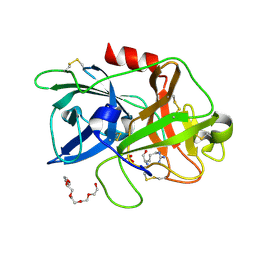
 | | The crystal structure of uPA complex with peptide inhibitor MH027 at pH4.6 | | 分子名称: | MH027, SULFATE ION, TETRAETHYLENE GLYCOL, ... | | 著者 | Jiang, L.G, Andreasen, P.A, Huang, M.D. | | 登録日 | 2010-09-21 | | 公開日 | 2011-08-10 | | 最終更新日 | 2023-11-01 | | 実験手法 | X-RAY DIFFRACTION (1.58 Å) | | 主引用文献 | The binding mechanism of a peptidic cyclic serine protease inhibitor
J.Mol.Biol., 412, 2011
|
|
4ZHL
 | |
3S87
 | | Structure of Yeast Ribonucleotide Reductase 1 with dGTP and ADP | | 分子名称: | 2'-DEOXYGUANOSINE-5'-TRIPHOSPHATE, ADENOSINE-5'-DIPHOSPHATE, MAGNESIUM ION, ... | | 著者 | Ahmad, M.F, Kaushal, P.S, Wan, Q, Wijeratna, S.R, Huang, M, Dealwis, C.D. | | 登録日 | 2011-05-27 | | 公開日 | 2012-04-11 | | 最終更新日 | 2023-09-13 | | 実験手法 | X-RAY DIFFRACTION (2.25 Å) | | 主引用文献 | Structural and biochemical basis of lethal mutant R293A of yeast ribonucleotide reductase
To be Published
|
|
4ZHM
 | |


