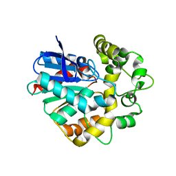
4C65
 
 | | Crystal structure of A. niger ochratoxinase | | 分子名称: | OCHRATOXINASE | | 著者 | Dobritzsch, D, Wang, H, Schneider, G, Yu, S. | | 登録日 | 2013-09-17 | | 公開日 | 2014-07-02 | | 最終更新日 | 2023-12-20 | | 実験手法 | X-RAY DIFFRACTION (2.2 Å) | | 主引用文献 | Structural and Functional Characterization of Ochratoxinase, a Novel Mycotoxin Degrading Enzyme.
Biochem.J., 462, 2014
|
|
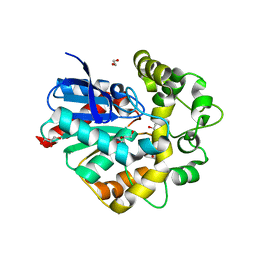
4C60
 | | Crystal structure of A. niger ochratoxinase | | 分子名称: | OCHRATOXINASE | | 著者 | Dobritzsch, D, Wang, H, Schneider, G, Yu, S. | | 登録日 | 2013-09-17 | | 公開日 | 2014-07-02 | | 最終更新日 | 2023-12-20 | | 実験手法 | X-RAY DIFFRACTION (2.5 Å) | | 主引用文献 | Structural and Functional Characterization of Ochratoxinase, a Novel Mycotoxin Degrading Enzyme.
Biochem.J., 462, 2014
|
|
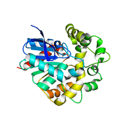
5O8H
 | | Crystal structure of R. ruber ADH-A, mutant Y294F, W295A, F43H, H39Y | | 分子名称: | Alcohol dehydrogenase, GLYCEROL, NICOTINAMIDE-ADENINE-DINUCLEOTIDE, ... | | 著者 | Dobritzsch, D, Maurer, D, Hamnevik, E, Reddy Enugala, T, Widersten, M. | | 登録日 | 2017-06-13 | | 公開日 | 2017-10-11 | | 最終更新日 | 2024-01-17 | | 実験手法 | X-RAY DIFFRACTION (1.96 Å) | | 主引用文献 | Relaxation of nonproductive binding and increased rate of coenzyme release in an alcohol dehydrogenase increases turnover with a nonpreferred alcohol enantiomer.
FEBS J., 284, 2017
|
|
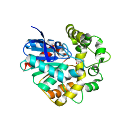
4C5Y
 | | Crystal structure of A. niger ochratoxinase | | 分子名称: | OCHRATOXINASE, ZINC ION | | 著者 | Dobritzsch, D, Wang, H, Schneider, G, Yu, S. | | 登録日 | 2013-09-17 | | 公開日 | 2014-07-02 | | 最終更新日 | 2023-12-20 | | 実験手法 | X-RAY DIFFRACTION (3 Å) | | 主引用文献 | Structural and Functional Characterization of Ochratoxinase, a Novel Mycotoxin Degrading Enzyme.
Biochem.J., 462, 2014
|
|
5MUB
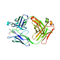 | | ACC1 Fab fragment in complex with citrullinated C1 epitope of CII (CG05) | | 分子名称: | ACC1 Fab fragment heavy chain, ACC1 Fab fragment light chain, triple-helical peptide containing the citrullinated C1 epitope of collagen type II,Collagen alpha-1(II) chain,triple-helical peptide containing the citrullinated C1 epitope of collagen type II | | 著者 | Dobritzsch, D, Holmdahl, R, Ge, C. | | 登録日 | 2017-01-13 | | 公開日 | 2017-07-19 | | 最終更新日 | 2024-01-17 | | 実験手法 | X-RAY DIFFRACTION (3.1 Å) | | 主引用文献 | Anti-citrullinated protein antibodies cause arthritis by cross-reactivity to joint cartilage.
JCI Insight, 2, 2017
|
|
5MU2
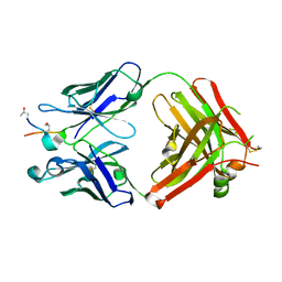 | | ACC1 Fab fragment in complex with CII583-591 (CG10) | | 分子名称: | ACC1 Fab fragment heavy chain, ACC1 Fab fragment light chain, synthetic peptide containing the CII583-591 epitope of collagen type II | | 著者 | Dobritzsch, D, Holmdahl, R, Ge, C. | | 登録日 | 2017-01-12 | | 公開日 | 2017-07-19 | | 最終更新日 | 2024-01-17 | | 実験手法 | X-RAY DIFFRACTION (2.7 Å) | | 主引用文献 | Anti-citrullinated protein antibodies cause arthritis by cross-reactivity to joint cartilage.
JCI Insight, 2, 2017
|
|
5MU0
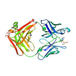 | | ACC1 Fab fragment in complex with citrullinated C1 epitope of CII (IA03) | | 分子名称: | IA03 peptide containing the citrullinated C1 epitope of collagen type II,Collagen alpha-1(II) chain,IA03 peptide containing the citrullinated C1 epitope of collagen type II, heavy chain of ACC1 antibody Fab fragment, light chain of ACC1 antibody Fab fragment | | 著者 | Dobritzsch, D, Holmdahl, R, Ge, C. | | 登録日 | 2017-01-11 | | 公開日 | 2017-07-19 | | 最終更新日 | 2024-01-17 | | 実験手法 | X-RAY DIFFRACTION (2.7 Å) | | 主引用文献 | Anti-citrullinated protein antibodies cause arthritis by cross-reactivity to joint cartilage.
JCI Insight, 2, 2017
|
|
5MV3
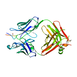 | | ACC1 Fab fragment in complex with CII583-591 (CG10) | | 分子名称: | heavy chain of ACC1 Fab fragment, light chain of ACC1 Fab fragment, synthetic peptide containing the CII583-591 epitope of collagen type II,Collagen alpha-1(II) chain,synthetic peptide containing the CII583-591 epitope of collagen type II, | | 著者 | Dobritzsch, D, Holmdahl, R, Ge, C. | | 登録日 | 2017-01-15 | | 公開日 | 2017-07-19 | | 最終更新日 | 2024-01-17 | | 実験手法 | X-RAY DIFFRACTION (2.95 Å) | | 主引用文献 | Anti-citrullinated protein antibodies cause arthritis by cross-reactivity to joint cartilage.
JCI Insight, 2, 2017
|
|
5MV4
 | | ACC1 Fab fragment in complex with citrullinated CII616-639 epitope of collagen type II (ptm23) | | 分子名称: | ACC1 antibody Fab fragment, heavy chain, light chain, ... | | 著者 | Dobritzsch, D, Holmdahl, R, Ge, C. | | 登録日 | 2017-01-15 | | 公開日 | 2017-07-19 | | 最終更新日 | 2024-01-17 | | 実験手法 | X-RAY DIFFRACTION (2.9 Å) | | 主引用文献 | Anti-citrullinated protein antibodies cause arthritis by cross-reactivity to joint cartilage.
JCI Insight, 2, 2017
|
|
5OD8
 | | Crystal structure of the RA-associated mAb B2 (Fab fragment) | | 分子名称: | B2 Fab fragment - Light chain, B2 Fab fragment - heavy chain | | 著者 | Dobritzsch, D, Ge, C, Holmdahl, R, Amara, K, Malmstrom, V. | | 登録日 | 2017-07-05 | | 公開日 | 2018-07-04 | | 最終更新日 | 2024-01-17 | | 実験手法 | X-RAY DIFFRACTION (2.2 Å) | | 主引用文献 | Structural Basis of Cross-Reactivity of Anti-Citrullinated Protein Antibodies.
Arthritis Rheumatol, 71, 2019
|
|
2Y5T
 | | Crystal structure of the pathogenic autoantibody CIIC1 in complex with the triple-helical C1 peptide | | 分子名称: | C1, CHLORIDE ION, CIIC1 FAB FRAGMENT HEAVY CHAIN, ... | | 著者 | Dobritzsch, D, Lindh, I, Schneider, N, Uysal, H, Nandakumar, K.S, Burkhardt, H, Schneider, G, Holmdahl, R. | | 登録日 | 2011-01-17 | | 公開日 | 2011-12-14 | | 最終更新日 | 2023-12-20 | | 実験手法 | X-RAY DIFFRACTION (2.2 Å) | | 主引用文献 | Crystal Structure of an Arthritogenic Anticollagen Immune Complex.
Arthritis Rheum., 63, 2011
|
|
7ZX4
 | | Clathrin N-terminal domain in complex with a HURP phospho-peptide | | 分子名称: | CHLORIDE ION, Clathrin heavy chain 1, Disks large-associated protein 5, ... | | 著者 | Kliche, J, Badgujar, D, Dobritzsch, D, Ivarsson, Y. | | 登録日 | 2022-05-20 | | 公開日 | 2023-03-29 | | 最終更新日 | 2024-02-07 | | 実験手法 | X-RAY DIFFRACTION (2.08 Å) | | 主引用文献 | Large-scale phosphomimetic screening identifies phospho-modulated motif-based protein interactions.
Mol.Syst.Biol., 19, 2023
|
|
6YUH
 | | Crystal structure of SMYD3 with diperodon R enantiomer bound to allosteric site | | 分子名称: | Diperodon, GLYCEROL, Histone-lysine N-methyltransferase SMYD3, ... | | 著者 | Cederfelt, D, Talibov, V.O, Dobritzsch, D, Danielson, U.H. | | 登録日 | 2020-04-27 | | 公開日 | 2021-01-13 | | 最終更新日 | 2024-01-24 | | 実験手法 | X-RAY DIFFRACTION (1.93 Å) | | 主引用文献 | Discovery of an Allosteric Ligand Binding Site in SMYD3 Lysine Methyltransferase.
Chembiochem, 22, 2021
|
|
8BBQ
 | | Determination of the structure of active tyrosinase from bacterium Verrucomicrobium spinosum | | 分子名称: | COPPER (II) ION, Core tyrosinase, GLYCEROL, ... | | 著者 | Fekry, M, Dave, K, Badgujar, D, Aurelius, O, Hamnevik, E, Dobritzsch, D, Danielson, H. | | 登録日 | 2022-10-14 | | 公開日 | 2023-09-20 | | 最終更新日 | 2023-10-11 | | 実験手法 | X-RAY DIFFRACTION (1.43 Å) | | 主引用文献 | The Crystal Structure of Tyrosinase from Verrucomicrobium spinosum Reveals It to Be an Atypical Bacterial Tyrosinase.
Biomolecules, 13, 2023
|
|
8BBR
 | | Determination of the structure of active tyrosinase from bacterium Verrucomicrobium spinosum | | 分子名称: | COPPER (II) ION, Core tyrosinase, SULFATE ION | | 著者 | Fekry, M, Dave, K, Badgujar, D, Aurelius, O, Hamnevik, E, Dobritzsch, D, Danielson, H. | | 登録日 | 2022-10-14 | | 公開日 | 2023-09-20 | | 最終更新日 | 2023-10-11 | | 実験手法 | X-RAY DIFFRACTION (1.64 Å) | | 主引用文献 | The Crystal Structure of Tyrosinase from Verrucomicrobium spinosum Reveals It to Be an Atypical Bacterial Tyrosinase.
Biomolecules, 13, 2023
|
|
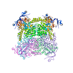
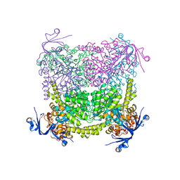
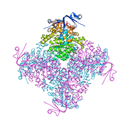
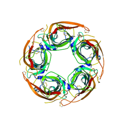
8PT4
 | | beta-Ureidopropionase tetramer | | 分子名称: | Beta-ureidopropionase | | 著者 | Cederfelt, D, Dobritzsch, D. | | 登録日 | 2023-07-13 | | 公開日 | 2024-01-10 | | 実験手法 | ELECTRON MICROSCOPY (3.33 Å) | | 主引用文献 | The Allosteric Regulation of Beta-Ureidopropionase Depends on Fine-Tuned Stability of Active-Site Loops and Subunit Interfaces.
Biomolecules, 13, 2023
|
|
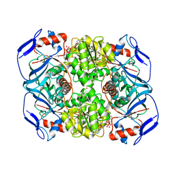
4UFP
 | | Laboratory evolved variant R-C1B1D33 of potato epoxide hydrolase StEH1 | | 分子名称: | EPOXIDE HYDROLASE | | 著者 | Carlsson, A.J, Bauer, P, Nilsson, M, Dobritzsch, D, Kamerlin, S.C.L, Widersten, M. | | 登録日 | 2015-03-17 | | 公開日 | 2016-04-13 | | 最終更新日 | 2023-12-20 | | 実験手法 | X-RAY DIFFRACTION (2.95 Å) | | 主引用文献 | Laboratory Evolved Enzymes Provide Snapshots of the Development of Enantioconvergence in Enzyme-Catalyzed Epoxide Hydrolysis.
Chembiochem, 17, 2016
|
|
4UHB
 | | Laboratory evolved variant R-C1 of potato epoxide hydrolase StEH1 | | 分子名称: | 1,2-ETHANEDIOL, EPOXIDE HYDROLASE, GLYCEROL | | 著者 | Nilsson, M.T.I, Carlsson, A.J, Dobritzsch, D, Widersten, M. | | 登録日 | 2015-03-23 | | 公開日 | 2016-04-13 | | 最終更新日 | 2024-01-10 | | 実験手法 | X-RAY DIFFRACTION (1.8 Å) | | 主引用文献 | Laboratory Evolved Enzymes Provide Snapshots of the Development of Enantioconvergence in Enzyme-Catalyzed Epoxide Hydrolysis.
Chembiochem, 17, 2016
|
|
4UFN
 | | Laboratory evolved variant R-C1B1 of potato epoxide hydrolase StEH1 | | 分子名称: | 1,4-DIETHYLENE DIOXIDE, EPOXIDE HYDROLASE | | 著者 | Carlsson, A.J, Bauer, P, Nilsson, M, Dobritzsch, D, Kamerlin, S.C.L, Widersten, M. | | 登録日 | 2015-03-17 | | 公開日 | 2016-04-13 | | 最終更新日 | 2023-12-20 | | 実験手法 | X-RAY DIFFRACTION (2 Å) | | 主引用文献 | Conformational Diversity and Enantioconvergence in Potato Epoxide Hydrolase 1.
Org.Biomol.Chem., 14, 2016
|
|
4UFO
 | | Laboratory evolved variant R-C1B1D33E6 of potato epoxide hydrolase StEH1 | | 分子名称: | EPOXIDE HYDROLASE | | 著者 | Carlsson, A.J, Bauer, P, Nilsson, M, Dobritzsch, D, Kamerlin, S.C.L, Widersten, M. | | 登録日 | 2015-03-17 | | 公開日 | 2016-04-13 | | 最終更新日 | 2023-12-20 | | 実験手法 | X-RAY DIFFRACTION (2.02 Å) | | 主引用文献 | Laboratory Evolved Enzymes Provide Snapshots of the Development of Enantioconvergence in Enzyme-Catalyzed Epoxide Hydrolysis.
Chembiochem, 17, 2016
|
|
8Q1T
 | | X-ray structure of acetylcholine binding protein (AChBP) in complex with IOTA739 | | 分子名称: | 1,10-PHENANTHROLINE, 2-acetamido-2-deoxy-beta-D-glucopyranose, Acetylcholine-binding protein, ... | | 著者 | Cederfelt, D, Lund, B.A, Boronat, P, Hennig, S, Dobritzsch, D, Danielson, U.H. | | 登録日 | 2023-08-01 | | 公開日 | 2024-06-05 | | 実験手法 | X-RAY DIFFRACTION (3 Å) | | 主引用文献 | Elucidating the regulation of ligand gated ion channels via biophysical studies of ligand-induced conformational dynamics of acetylcholine binding proteins
To Be Published
|
|
1ZPD
 | | PYRUVATE DECARBOXYLASE FROM ZYMOMONAS MOBILIS | | 分子名称: | CITRIC ACID, MAGNESIUM ION, MONO-{4-[(4-AMINO-2-METHYL-PYRIMIDIN-5-YLMETHYL)-AMINO]-2-HYDROXY-3-MERCAPTO-PENT-3-ENYL-PHOSPHONO} ESTER, ... | | 著者 | Lu, G, Dobritzsch, D, Schneider, G. | | 登録日 | 1998-04-17 | | 公開日 | 1999-02-02 | | 最終更新日 | 2024-05-22 | | 実験手法 | X-RAY DIFFRACTION (1.86 Å) | | 主引用文献 | High resolution crystal structure of pyruvate decarboxylase from Zymomonas mobilis. Implications for substrate activation in pyruvate decarboxylases.
J.Biol.Chem., 273, 1998
|
|
7NDP
 | | X-ray structure of acetylcholine-binding protein (AChBP) in complex with FL001856. | | 分子名称: | 2-acetamido-2-deoxy-beta-D-glucopyranose, 2-acetamido-2-deoxy-beta-D-glucopyranose-(1-4)-2-acetamido-2-deoxy-beta-D-glucopyranose, 6-bromanylspiro[3~{H}-chromene-2,4'-piperidine]-4-one, ... | | 著者 | Cederfelt, D, Boronat, P, Dobritzsch, D, Hennig, S, Fitzgerald, E.A, de Esch, I.J.P, Danielson, U.H. | | 登録日 | 2021-02-02 | | 公開日 | 2021-04-07 | | 最終更新日 | 2024-01-31 | | 実験手法 | X-RAY DIFFRACTION (2 Å) | | 主引用文献 | Discovery of fragments inducing conformational effects in dynamic proteins using a second-harmonic generation biosensor.
Rsc Adv, 11, 2021
|
|
7NDV
 | | X-ray structure of acetylcholine-binding protein (AChBP) in complex with FL001888. | | 分子名称: | 2-acetamido-2-deoxy-beta-D-glucopyranose, 4-[4-(trifluoromethyl)phenoxy]piperidine, Acetylcholine-binding protein, ... | | 著者 | Cederfelt, D, Boronat, P, Dobritzsch, D, Hennig, S, Fitzgerald, E.A, de Esch, I.J.P, Danielson, U.H. | | 登録日 | 2021-02-02 | | 公開日 | 2021-04-07 | | 最終更新日 | 2024-01-31 | | 実験手法 | X-RAY DIFFRACTION (1.7 Å) | | 主引用文献 | Discovery of fragments inducing conformational effects in dynamic proteins using a second-harmonic generation biosensor
RSC Advances, 11, 2021
|
|
7NZF
 | | Crystal structure of HLA-DR4 in complex with a mutated human collagen type II peptide | | 分子名称: | 2-acetamido-2-deoxy-beta-D-glucopyranose, HLA class II histocompatibility antigen, DR alpha chain, ... | | 著者 | Ge, C, Dobritzsch, D, Holmdahl, R. | | 登録日 | 2021-03-24 | | 公開日 | 2022-05-11 | | 最終更新日 | 2024-01-31 | | 実験手法 | X-RAY DIFFRACTION (1.9 Å) | | 主引用文献 | Key interactions in the trimolecular complex consisting of the rheumatoid arthritis-associated DRB1*04:01 molecule, the major glycosylated collagen II peptide and the T-cell receptor.
Ann Rheum Dis, 81, 2022
|
|


