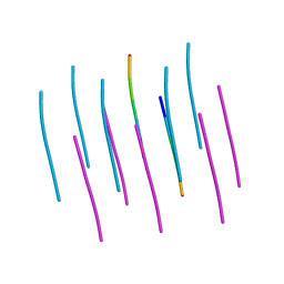
6CFH
 
 | |
6CB9
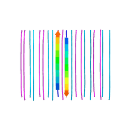
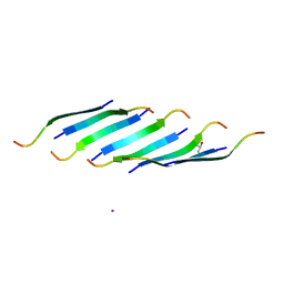
 | | Segment AALQSS from the low complexity domain of TDP-43, residues 328-333 | | 分子名称: | AALQSS | | 著者 | Guenther, E.L, Cao, Q, Lu, J, Sawaya, M.R, Eisenberg, D.S. | | 登録日 | 2018-02-02 | | 公開日 | 2018-04-18 | | 最終更新日 | 2024-03-13 | | 実験手法 | X-RAY DIFFRACTION (1.1 Å) | | 主引用文献 | Atomic structures of TDP-43 LCD segments and insights into reversible or pathogenic aggregation.
Nat. Struct. Mol. Biol., 25, 2018
|
|
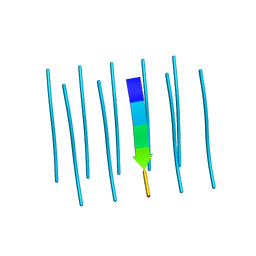
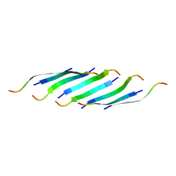
6CEW
 | | Segment AMMAAA from the low complexity domain of TDP-43, residues 321-326 | | 分子名称: | AMMAAA | | 著者 | Guenther, E.L, Cao, Q, Lu, J, Sawaya, M.R, Eisenberg, D.S. | | 登録日 | 2018-02-12 | | 公開日 | 2018-04-18 | | 最終更新日 | 2024-04-03 | | 実験手法 | X-RAY DIFFRACTION (1.2 Å) | | 主引用文献 | Atomic structures of TDP-43 LCD segments and insights into reversible or pathogenic aggregation.
Nat. Struct. Mol. Biol., 25, 2018
|
|
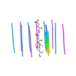
6CF4
 | | Segment NFGTFS, with familial mutation A315T and phosphorylated threonine, from the low complexity domain of TDP-43, residues 312-317 | | 分子名称: | NFGTFS | | 著者 | Guenther, E.L, Cao, Q, Boyer, D.R, Sawaya, M.R, Eisenberg, D.S. | | 登録日 | 2018-02-13 | | 公開日 | 2018-05-23 | | 最終更新日 | 2021-06-30 | | 実験手法 | ELECTRON CRYSTALLOGRAPHY (0.75 Å) | | 主引用文献 | Atomic structures of TDP-43 LCD segments and insights into reversible or pathogenic aggregation.
Nat. Struct. Mol. Biol., 25, 2018
|
|
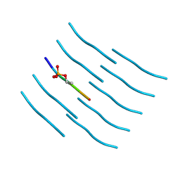
5WIA
 | | Crystal structure of the segment, GNNSYS, from the low complexity domain of TDP-43, residues 370-375 | | 分子名称: | TAR DNA-binding protein 43 | | 著者 | Guenther, E.L, Trinh, H, Sawaya, M.R, Eisenberg, D.S. | | 登録日 | 2017-07-18 | | 公開日 | 2018-04-25 | | 最終更新日 | 2024-04-03 | | 実験手法 | X-RAY DIFFRACTION (1.002 Å) | | 主引用文献 | Atomic structures of TDP-43 LCD segments and insights into reversible or pathogenic aggregation.
Nat. Struct. Mol. Biol., 25, 2018
|
|
5WKD
 | | Crystal structure of the segment, GNNQGSN, from the low complexity domain of TDP-43, residues 300-306 | | 分子名称: | TAR DNA-binding protein 43 | | 著者 | Guenther, E.L, Trinh, H, Sawaya, M.R, Cascio, D, Eisenberg, D.S. | | 登録日 | 2017-07-25 | | 公開日 | 2018-04-18 | | 最終更新日 | 2024-04-03 | | 実験手法 | X-RAY DIFFRACTION (1.8 Å) | | 主引用文献 | Atomic structures of TDP-43 LCD segments and insights into reversible or pathogenic aggregation.
Nat. Struct. Mol. Biol., 25, 2018
|
|
5WHP
 | | Crystal structure of the segment, NFGTFS, from the A315T familial variant of the low complexity domain of TDP-43, residues 312-317 | | 分子名称: | Segment of TAR DNA-binding protein 43 | | 著者 | Guenther, E.L, Sawaya, M.R, Eisenberg, D.S. | | 登録日 | 2017-07-17 | | 公開日 | 2018-05-23 | | 最終更新日 | 2024-03-13 | | 実験手法 | X-RAY DIFFRACTION (1 Å) | | 主引用文献 | Atomic structures of TDP-43 LCD segments and insights into reversible or pathogenic aggregation.
Nat. Struct. Mol. Biol., 25, 2018
|
|
5WIQ
 | | Crystal structure of the segment, GFNGGFG, from the low complexity domain of TDP-43, residues 396-402 | | 分子名称: | TAR DNA-binding protein 43 | | 著者 | Guenther, E.L, Sawaya, M.R, Eisenberg, D.S. | | 登録日 | 2017-07-19 | | 公開日 | 2018-04-18 | | 最終更新日 | 2023-10-04 | | 実験手法 | X-RAY DIFFRACTION (1.25 Å) | | 主引用文献 | Atomic structures of TDP-43 LCD segments and insights into reversible or pathogenic aggregation.
Nat. Struct. Mol. Biol., 25, 2018
|
|
5W50
 | | Crystal structure of the segment, LIIKGI, from the RRM2 of TDP-43, residues 248-253 | | 分子名称: | TAR DNA-binding protein 43 | | 著者 | Guenther, E.L, Trinh, H, Sawaya, M.R, Eisenberg, D.S. | | 登録日 | 2017-06-13 | | 公開日 | 2018-02-21 | | 最終更新日 | 2023-10-04 | | 実験手法 | X-RAY DIFFRACTION (1.4 Å) | | 主引用文献 | Atomic-level evidence for packing and positional amyloid polymorphism by segment from TDP-43 RRM2.
Nat. Struct. Mol. Biol., 25, 2018
|
|
5W52
 | | MicroED structure of the segment, DLIIKGISVHI, from the RRM2 of TDP-43, residues 247-257 | | 分子名称: | TAR DNA-binding protein 43 | | 著者 | Guenther, E.L, Sawaya, M.R, Cascio, D, Eisenberg, D.S. | | 登録日 | 2017-06-13 | | 公開日 | 2018-02-21 | | 最終更新日 | 2024-04-03 | | 実験手法 | ELECTRON CRYSTALLOGRAPHY (1.4 Å) | | 主引用文献 | Atomic-level evidence for packing and positional amyloid polymorphism by segment from TDP-43 RRM2.
Nat. Struct. Mol. Biol., 25, 2018
|
|
5WKB
 | | MicroED structure of the segment, NFGEFS, from the A315E familial variant of the low complexity domain of TDP-43, residues 312-317 | | 分子名称: | TAR DNA-binding protein 43 | | 著者 | Guenther, E.L, Sawaya, M.R, Cascio, D, Eisenberg, D.S. | | 登録日 | 2017-07-24 | | 公開日 | 2018-05-23 | | 最終更新日 | 2024-03-13 | | 実験手法 | ELECTRON CRYSTALLOGRAPHY (1 Å) | | 主引用文献 | Atomic structures of TDP-43 LCD segments and insights into reversible or pathogenic aggregation.
Nat. Struct. Mol. Biol., 25, 2018
|
|
5W7V
 | |
5WHN
 | | Crystal structure of the segment, NFGAFS, from the low complexity domain of TDP-43, residues 312-317 | | 分子名称: | Segment of TAR DNA-binding protein 43 | | 著者 | Guenther, E.L, Sawaya, M.R, Eisenberg, D.S. | | 登録日 | 2017-07-17 | | 公開日 | 2018-04-25 | | 最終更新日 | 2023-10-04 | | 実験手法 | X-RAY DIFFRACTION (1.1 Å) | | 主引用文献 | Atomic structures of TDP-43 LCD segments and insights into reversible or pathogenic aggregation.
Nat. Struct. Mol. Biol., 25, 2018
|
|
4RIK
 | | Amyloid forming segment, AVVTGVTAV, from the NAC domain of Parkinson's disease protein alpha-synuclein, residues 69-77 | | 分子名称: | Alpha-synuclein | | 著者 | Guenther, E.L, Sawaya, M.R, Ivanova, M, Eisenberg, D.S. | | 登録日 | 2014-10-06 | | 公開日 | 2015-08-26 | | 最終更新日 | 2024-04-03 | | 実験手法 | X-RAY DIFFRACTION (1.854 Å) | | 主引用文献 | Structure of the toxic core of alpha-synuclein from invisible crystals.
Nature, 525, 2015
|
|
4RIL
 | | Structure of the amyloid forming segment, GAVVTGVTAVA, from the NAC domain of Parkinson's disease protein alpha-synuclein, residues 68-78, determined by electron diffraction | | 分子名称: | Alpha-synuclein | | 著者 | Rodriguez, J.A, Ivanova, M, Sawaya, M.R, Cascio, D, Reyes, F, Shi, D, Johnson, L, Guenther, E, Sangwan, S, Hattne, J, Nannenga, B, Brewster, A.S, Messerschmidt, M, Boutet, S, Sauter, N.K, Gonen, T, Eisenberg, D.S. | | 登録日 | 2014-10-06 | | 公開日 | 2015-08-26 | | 最終更新日 | 2023-09-20 | | 実験手法 | ELECTRON CRYSTALLOGRAPHY (1.43 Å) | | 主引用文献 | Structure of the toxic core of alpha-synuclein from invisible crystals.
Nature, 525, 2015
|
|
4ZNN
 | | MicroED structure of the segment, GVVHGVTTVA, from the A53T familial mutant of Parkinson's disease protein, alpha-synuclein residues 47-56 | | 分子名称: | Alpha-synuclein | | 著者 | Rodriguez, J.A, Ivanova, M, Sawaya, M.R, Cascio, D, Reyes, F, Shi, D, Johnson, L, Guenther, E, Sangwan, S, Hattne, J, Nannenga, B, Brewster, A.S, Messerschmidt, M, Boutet, S, Sauter, N.K, Gonen, T, Eisenberg, D.S. | | 登録日 | 2015-05-05 | | 公開日 | 2015-09-09 | | 最終更新日 | 2024-03-06 | | 実験手法 | ELECTRON CRYSTALLOGRAPHY (1.41 Å) | | 主引用文献 | Structure of the toxic core of alpha-synuclein from invisible crystals.
Nature, 525, 2015
|
|
5DLI
 | | Corkscrew assembly of SOD1 residues 28-38 | | 分子名称: | GLYCEROL, IODIDE ION, Superoxide dismutase [Cu-Zn] | | 著者 | Sangwan, S, Zhao, A, Sawaya, M.R, Eisenberg, D. | | 登録日 | 2015-09-05 | | 公開日 | 2016-09-14 | | 最終更新日 | 2024-03-06 | | 実験手法 | X-RAY DIFFRACTION (2.1 Å) | | 主引用文献 | Atomic structure of a toxic, oligomeric segment of SOD1 linked to amyotrophic lateral sclerosis (ALS).
Proc. Natl. Acad. Sci. U.S.A., 114, 2017
|
|
5IIW
 | | Corkscrew assembly of SOD1 residues 28-38 without potassium iodide | | 分子名称: | Superoxide dismutase [Cu-Zn] | | 著者 | Sangwan, S, Zhao, A, Sawaya, M.R, Eisenberg, D. | | 登録日 | 2016-03-01 | | 公開日 | 2017-06-28 | | 最終更新日 | 2024-03-06 | | 実験手法 | X-RAY DIFFRACTION (2 Å) | | 主引用文献 | Atomic structure of a toxic, oligomeric segment of SOD1 linked to amyotrophic lateral sclerosis (ALS).
Proc. Natl. Acad. Sci. U.S.A., 114, 2017
|
|
4NIP
 | |
4NIN
 | |
4NIO
 | |
3I58
 | |
3I53
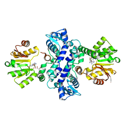 | |
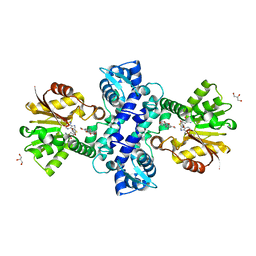
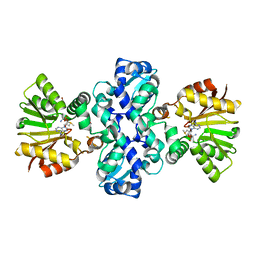
3I64
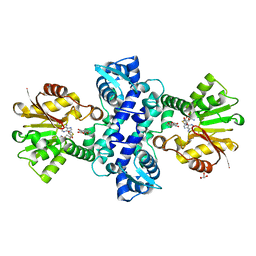 | | Crystal structure of an O-methyltransferase (NcsB1) from neocarzinostatin biosynthesis in complex with S-adenosyl-L-homocysteine (SAH) and 1,4-dihydroxy-2-naphthoic acid (DHN) | | 分子名称: | 1,4-dihydroxy-2-naphthoic acid, GLYCEROL, O-methyltransferase, ... | | 著者 | Cooke, H.A, Bruner, S.D. | | 登録日 | 2009-07-06 | | 公開日 | 2009-09-01 | | 最終更新日 | 2023-09-06 | | 実験手法 | X-RAY DIFFRACTION (3 Å) | | 主引用文献 | Molecular basis of substrate promiscuity for the SAM-dependent O-methyltransferase NcsB1, involved in the biosynthesis of the enediyne antitumor antibiotic neocarzinostatin.
Biochemistry, 48, 2009
|
|
3I5U
 | |


