 Movie
Movie Controller
 Structure viewers
 About EMN search


-Search query
-Search result
Showing 1 - 50 of 122 items for (author: le & ttn)
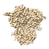
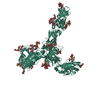
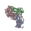
EMDB-41363: 
Cryo-EM structure of DDB1deltaB-DDA1-DCAF5

PDB-8tl6:
Cryo-EM structure of DDB1deltaB-DDA1-DCAF5

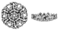
PDB-8qox:
Two-component assembly of SlaA and SlaB S-layer proteins of Sulfolobus acidocaldarius

PDB-8qp0:
A hexamer pore in the S-layer of Sulfolobus acidocaldarius formed by SlaA protein
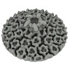
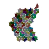
EMDB-18127:
S-layer of archaeon Sulfolobus acidocaldarius by subtomogram averaging
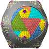
EMDB-19035:
Composite map of the Emiliania huxleyi virus 201 (EhV-201) symmetry expanded from cryo-EM structure of virion vertex 120 nm in diameter.
EMDB-19036:
Cryo-EM structure of the Emiliania huxleyi virus 201 (EhV-201) virion vertex with a diameter of 50 nm and a mask applied on the capsid layer.
PDB-8rbs:
Emiliania huxleyi virus 201 (EhV-201) asymmetrical unit of capsid proteins predicted by AlphaFold2 fitted into the cryo-EM density of EhV-201 virion composite map.
PDB-8rbt:
Emiliania huxleyi virus 201 (EhV-201) capsid proteins predicted by AlphaFold2 fitted into a cryo-EM density map of the EhV-201 virion capsid.

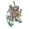
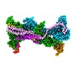
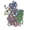
EMDB-29578:
G4 RNA-mediated PRC2 dimer
EMDB-29647:
Body1 of G4 RNA-mediated PRC2 dimer from multibody refinement
EMDB-29656:
Body2 of G4 RNA-mediated PRC2 dimer from multibody refinement

PDB-8fyh:
G4 RNA-mediated PRC2 dimer

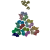
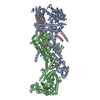
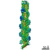
EMDB-15530:
S-layer protein SlaA from Sulfolobus acidocaldarius at pH 10.0

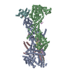
EMDB-15531:
S-layer protein SlaA from Sulfolobus acidocaldarius at pH 7.0

PDB-8an2:
S-layer protein SlaA from Sulfolobus acidocaldarius at pH 10.0

PDB-8an3:
S-layer protein SlaA from Sulfolobus acidocaldarius at pH 7.0

EMDB-17823:
Tomogram of the Emiliania huxleyi virus 201 (EhV-201) purified particles used for subtomogram averaging.

EMDB-17649:
Cryo-EM structure of the Emiliania huxleyi virus 201 (EhV-201) virion vertex with a diameter of 50 nm.

EMDB-17650:
Cryo-EM structure of the Emiliania huxleyi virus 201 (EhV-201) virion vertex with a diameter of 50 nm.

EMDB-17651:
Cryo-EM structure of the Emiliania huxleyi virus 201 (EhV-201) virion vertex with a diameter of 120 nm.

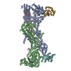
EMDB-14635:
S-layer protein SlaA from Sulfolobus acidocaldarius at pH 4.0
PDB-7zcx:
S-layer protein SlaA from Sulfolobus acidocaldarius at pH 4.0

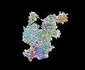
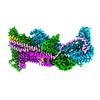
EMDB-27777:
Streptomyces venezuelae RNAP transcription open promoter complex with WhiA and WhiB transcription factors

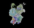
EMDB-27778:
Streptomyces venezuelae RNAP unconstrained open promoter complex with WhiA and WhiB transcription factors

PDB-8dy7:
Streptomyces venezuelae RNAP transcription open promoter complex with WhiA and WhiB transcription factors
PDB-8dy9:
Streptomyces venezuelae RNAP unconstrained open promoter complex with WhiA and WhiB transcription factors

EMDB-15136:
In-cell structure of the actin filament Arp2/3 complex branch junction in lamellipodia of WT B16-F1 mouse melanoma cells

EMDB-15137:
Tomogram of a lamellipodium of a WT B16-F1 mouse melanoma cell

EMDB-15138:
In-cell structure of the actin filament Arp2/3 complex branch junction in lamellipodia of ArpC5 knockout B16-F1 mouse melanoma cells

EMDB-15139:
Tomogram of a lamellipodium of an ArpC5 knockout B16-F1 mouse melanoma cell

EMDB-15140:
In-cell structure of the actin filament Arp2/3 complex branch junction in lamellipodia of ArpC5L knockout B16-F1 mouse melanoma cells

EMDB-15141:
Tomogram of a lamellipodium of an ArpC5L knockout B16-F1 mouse melanoma cell
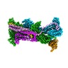
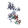
EMDB-26732:
Cryo-EM structure of WAVE Regulatory Complex
EMDB-26733:
Cryo-EM structure of D-site Rac1-bound WAVE Regulatory Complex
EMDB-26734:
Cryo-EM structure of WAVE regulatory complex with Rac1 bound on both A and D site
PDB-7usc:
Cryo-EM structure of WAVE Regulatory Complex
PDB-7usd:
Cryo-EM structure of D-site Rac1-bound WAVE Regulatory Complex
PDB-7use:
Cryo-EM structure of WAVE regulatory complex with Rac1 bound on both A and D site
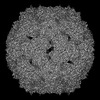
EMDB-14183:
Cryo-EM structure of coxsackievirus A6 altered particle
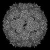
EMDB-14184:
Cryo-EM structure of coxsackievirus A6 empty particle
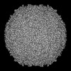
EMDB-14186:
Cryo-EM structure of coxsackievirus A6 mature virion
PDB-7qvx:
Cryo-EM structure of coxsackievirus A6 altered particle
PDB-7qvy:
Cryo-EM structure of coxsackievirus A6 empty particle
PDB-7qw9:
Cryo-EM structure of coxsackievirus A6 mature virion

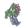
EMDB-14591:
CryoEM structure of SARS-CoV-2 spike monomer in complex with neutralising antibody P008_60
PDB-7zbu:
CryoEM structure of SARS-CoV-2 spike monomer in complex with neutralising antibody P008_60
EMDB-13343:
Structure of capping protein bound to the barbed end of a cytoplasmic actin filament
PDB-7pdz:
Structure of capping protein bound to the barbed end of a cytoplasmic actin filament

EMDB-10847:
CIII2/CIV respiratory supercomplex from Saccharomyces cerevisiae
Pages:
 wwPDB to switch to version 3 of the EMDB data model
