 Movie
Movie Controller


+ Open data

- Basic information
| Entry |  | |||||||||
|---|---|---|---|---|---|---|---|---|---|---|
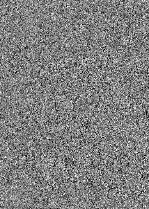
| Title | Tomogram of a lamellipodium of a WT B16-F1 mouse melanoma cell | |||||||||
 Map data Map data | Tomogram of a lamellipodium of a WT B16-F1 mouse melanoma cell | |||||||||
 Sample Sample |
| |||||||||
| Biological species |  | |||||||||
| Method | electron tomography / cryo EM | |||||||||
 Authors Authors | Faessler F / Javoor MG / Datler J / Doering H / Hofer FW / Dimchev G / Hodirnau VV / Rottner K / Schur FKM | |||||||||
| Funding support |  Austria, 1 items Austria, 1 items
| |||||||||
 Citation Citation | Journal: Sci Adv / Year: 2023 Title: ArpC5 isoforms regulate Arp2/3 complex-dependent protrusion through differential Ena/VASP positioning. Authors: Florian Fäßler / Manjunath G Javoor / Julia Datler / Hermann Döring / Florian W Hofer / Georgi Dimchev / Victor-Valentin Hodirnau / Jan Faix / Klemens Rottner / Florian K M Schur /  Abstract: Regulation of the Arp2/3 complex is required for productive nucleation of branched actin networks. An emerging aspect of regulation is the incorporation of subunit isoforms into the Arp2/3 complex. ...Regulation of the Arp2/3 complex is required for productive nucleation of branched actin networks. An emerging aspect of regulation is the incorporation of subunit isoforms into the Arp2/3 complex. Specifically, both ArpC5 subunit isoforms, ArpC5 and ArpC5L, have been reported to fine-tune nucleation activity and branch junction stability. We have combined reverse genetics and cellular structural biology to describe how ArpC5 and ArpC5L differentially affect cell migration. Both define the structural stability of ArpC1 in branch junctions and, in turn, by determining protrusion characteristics, affect protein dynamics and actin network ultrastructure. ArpC5 isoforms also affect the positioning of members of the Ena/Vasodilator-stimulated phosphoprotein (VASP) family of actin filament elongators, which mediate ArpC5 isoform-specific effects on the actin assembly level. Our results suggest that ArpC5 and Ena/VASP proteins are part of a signaling pathway enhancing cell migration. | |||||||||
| History |
|
- Structure visualization
| Supplemental images |
|---|

- Downloads & links
-EMDB archive
| Map data | emd_15137.map.gz | 260.6 MB |  EMDB map data format EMDB map data format | |
|---|---|---|---|---|
| Header (meta data) | emd-15137-v30.xmlemd-15137.xml | 12.2 KB 12.2 KB | Display Display | EMDB header |
| Images | 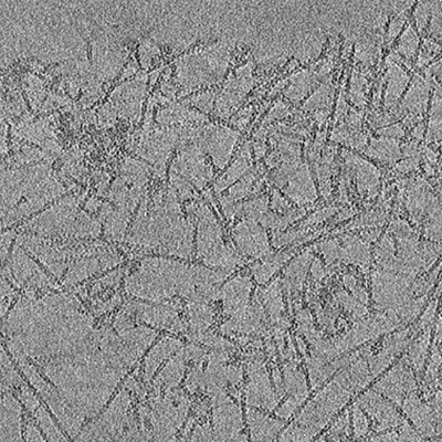 emd_15137.png emd_15137.png | 240.7 KB | ||
| Archive directory |  http://ftp.pdbj.org/pub/emdb/structures/EMD-15137ftp://ftp.pdbj.org/pub/emdb/structures/EMD-15137 http://ftp.pdbj.org/pub/emdb/structures/EMD-15137ftp://ftp.pdbj.org/pub/emdb/structures/EMD-15137 | HTTPS FTP |
-Related structure data


-Links
| EMDB pages | EMDB (EBI/PDBe) / EMDataResource |
|---|
-Map
| File | Download / File: emd_15137.map.gz / Format: CCP4 / Size: 281.3 MB / Type: IMAGE STORED AS FLOATING POINT NUMBER (4 BYTES) | ||||||||||||||||||||||||||||||||
|---|---|---|---|---|---|---|---|---|---|---|---|---|---|---|---|---|---|---|---|---|---|---|---|---|---|---|---|---|---|---|---|---|---|
| Annotation | Tomogram of a lamellipodium of a WT B16-F1 mouse melanoma cell | ||||||||||||||||||||||||||||||||
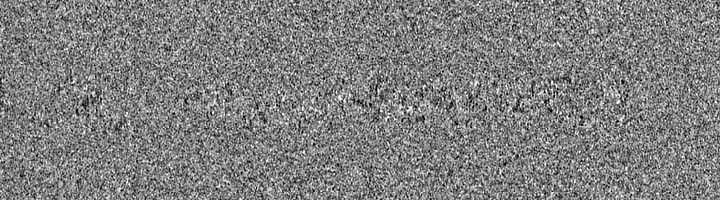
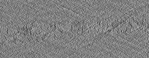
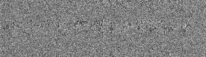
| Projections & slices | Image control
Images are generated by Spider. generated in cubic-lattice coordinate | ||||||||||||||||||||||||||||||||
| Voxel size | X=Y=Z: 13.544 Å | ||||||||||||||||||||||||||||||||
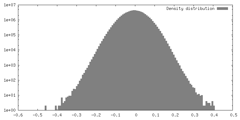
| Density |
| ||||||||||||||||||||||||||||||||
| Symmetry | Space group: 1 | ||||||||||||||||||||||||||||||||
| Details | EMDB XML:
|
 Z (Sec.)
 Y (Row.)
 X (Col.)


-Supplemental data
- Sample components
-Entire : Tomogram of a lamellipodium of a WT B16-F1 mouse melanoma cell
| Entire | Name: Tomogram of a lamellipodium of a WT B16-F1 mouse melanoma cell |
|---|---|
| Components |
|
-Supramolecule #1: Tomogram of a lamellipodium of a WT B16-F1 mouse melanoma cell
| Supramolecule | Name: Tomogram of a lamellipodium of a WT B16-F1 mouse melanoma cell type: cell / ID: 1 / Parent: 0 |
|---|---|
| Source (natural) | Organism: |
-Experimental details
-Structure determination
| Method | cryo EM |
|---|---|
 Processing Processing | electron tomography |
| Aggregation state | cell |
-Sample preparation
| Buffer | pH: 6.2 Component:
Details: Adjust to pH 6.2 using NaOH Immediately prior to use, add Phalloidin to a final concentration of 1ug/ml | ||||||||||||||
|---|---|---|---|---|---|---|---|---|---|---|---|---|---|---|---|
| Grid | Model: Quantifoil R2/2 / Material: GOLD / Mesh: 200 / Support film - Material: CARBON / Support film - topology: HOLEY / Pretreatment - Type: GLOW DISCHARGE / Pretreatment - Time: 120 sec. / Pretreatment - Atmosphere: AIR Details: After glow discharging of the grid and prior to the seeding of cells, the grid was coated using 25ug/ml Laminin for 60min | ||||||||||||||
| Vitrification | Cryogen name: ETHANE / Chamber humidity: 80 % / Chamber temperature: 277 K / Instrument: LEICA EM GP | ||||||||||||||
| Sectioning | Other: NO SECTIONING | ||||||||||||||
| Fiducial marker | Manufacturer: Aurion / Diameter: 10 nm |
- Electron microscopy
| Microscope | FEI TITAN KRIOS |
|---|---|
| Specialist optics | Energy filter - Name: GIF Bioquantum / Energy filter - Slit width: 20 eV |
| Image recording | Film or detector model: GATAN K3 (6k x 4k) / Number grids imaged: 1 / Number real images: 67 / Average electron dose: 2.79 e/Å2 Details: Images were collected in movie-mode with 8 frames per tilt |
| Electron beam | Acceleration voltage: 300 kV / Electron source:  FIELD EMISSION GUN FIELD EMISSION GUN |
| Electron optics | C2 aperture diameter: 50.0 µm / Illumination mode: FLOOD BEAM / Imaging mode: BRIGHT FIELD / Cs: 2.7 mm / Nominal defocus max: 4.5 µm / Nominal defocus min: 1.5 µm / Nominal magnification: 53000 |
| Sample stage | Specimen holder model: FEI TITAN KRIOS AUTOGRID HOLDER / Cooling holder cryogen: NITROGEN |
| Experimental equipment |  Model: Titan Krios / Image courtesy: FEI Company |
-Image processing
| Final reconstruction | Algorithm: BACK PROJECTION / Software - Name: RELION (ver. 3.0.8) / Number images used: 65 |
|---|
