 Movie
Movie Controller


+ Open data

- Basic information
| Entry | 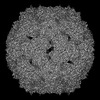 | |||||||||
|---|---|---|---|---|---|---|---|---|---|---|
| Title | Cryo-EM structure of coxsackievirus A6 altered particle | |||||||||
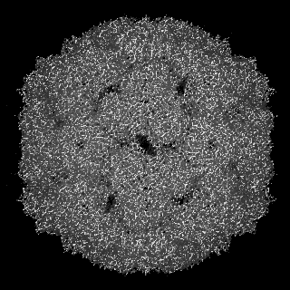
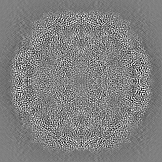
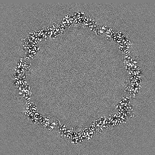
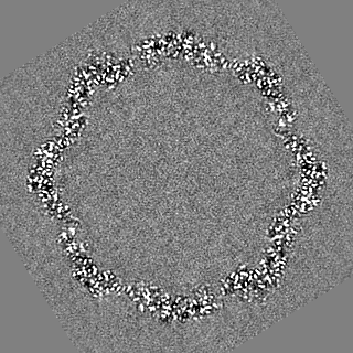
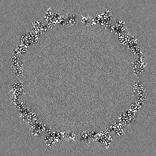
 Map data Map data | Sharpened map of CV-A6 altered particle at 2.5 A resolution | |||||||||
 Sample Sample |
| |||||||||
 Keywords Keywords | enterovirus / coxsackievirus A6 / altered particle / capsid / cryo-EM / VIRUS | |||||||||
| Function / homology |  Function and homology information Function and homology informationsymbiont-mediated suppression of host cytoplasmic pattern recognition receptor signaling pathway via inhibition of MDA-5 activity / picornain 2A / symbiont-mediated suppression of host mRNA export from nucleus / symbiont genome entry into host cell via pore formation in plasma membrane / picornain 3C / T=pseudo3 icosahedral viral capsid / host cell cytoplasmic vesicle membrane / ribonucleoside triphosphate phosphatase activity / nucleoside-triphosphate phosphatase / channel activity ...symbiont-mediated suppression of host cytoplasmic pattern recognition receptor signaling pathway via inhibition of MDA-5 activity / picornain 2A / symbiont-mediated suppression of host mRNA export from nucleus / symbiont genome entry into host cell via pore formation in plasma membrane / picornain 3C / T=pseudo3 icosahedral viral capsid / host cell cytoplasmic vesicle membrane / ribonucleoside triphosphate phosphatase activity / nucleoside-triphosphate phosphatase / channel activity / monoatomic ion transmembrane transport / DNA replication / RNA helicase activity / endocytosis involved in viral entry into host cell / symbiont-mediated activation of host autophagy / RNA-directed RNA polymerase / cysteine-type endopeptidase activity / viral RNA genome replication / RNA-directed RNA polymerase activity / DNA-templated transcription / virion attachment to host cell / host cell nucleus / structural molecule activity / proteolysis / RNA binding / zinc ion binding / ATP binding Similarity search - Function | |||||||||
| Biological species |  Coxsackievirus A6 Coxsackievirus A6 | |||||||||
| Method | single particle reconstruction / cryo EM / Resolution: 2.5 Å | |||||||||
 Authors Authors | Buttner CR / Plevka P | |||||||||
| Funding support |  Czech Republic, 2 items Czech Republic, 2 items
| |||||||||
 Citation Citation | Journal: Commun Biol / Year: 2022 Title: Cryo-electron microscopy and image classification reveal the existence and structure of the coxsackievirus A6 virion. Authors: Carina R Büttner / Radovan Spurný / Tibor Füzik / Pavel Plevka / Abstract: Coxsackievirus A6 (CV-A6) has recently overtaken enterovirus A71 and CV-A16 as the primary causative agent of hand, foot, and mouth disease worldwide. Virions of CV-A6 were not identified in previous ...Coxsackievirus A6 (CV-A6) has recently overtaken enterovirus A71 and CV-A16 as the primary causative agent of hand, foot, and mouth disease worldwide. Virions of CV-A6 were not identified in previous structural studies, and it was speculated that the virus is unique among enteroviruses in using altered particles with expanded capsids to infect cells. In contrast, the virions of other enteroviruses are required for infection. Here we used cryo-electron microscopy (cryo-EM) to determine the structures of the CV-A6 virion, altered particle, and empty capsid. We show that the CV-A6 virion has features characteristic of virions of other enteroviruses, including a compact capsid, VP4 attached to the inner capsid surface, and fatty acid-like molecules occupying the hydrophobic pockets in VP1 subunits. Furthermore, we found that in a purified sample of CV-A6, the ratio of infectious units to virions is 1 to 500. Therefore, it is likely that virions of CV-A6 initiate infection, like those of other enteroviruses. Our results provide evidence that future vaccines against CV-A6 should target its virions instead of the antigenically distinct altered particles. Furthermore, the structure of the virion provides the basis for the rational development of capsid-binding inhibitors that block the genome release of CV-A6. | |||||||||
| History |
|
- Structure visualization
| Supplemental images |
|---|

- Downloads & links
-EMDB archive
| Map data | emd_14183.map.gz | 98.3 MB | EMDB map data format | |
|---|---|---|---|---|
| Header (meta data) | emd-14183-v30.xmlemd-14183.xml | 26.4 KB 26.4 KB | Display Display | EMDB header |
| FSC (resolution estimation) | emd_14183_fsc.xml | 18 KB | Display | FSC data file |
| Images | 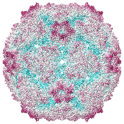 emd_14183.png emd_14183.png | 327.9 KB | ||
| Filedesc metadata | emd-14183.cif.gz | 7 KB | ||
| Others | emd_14183_additional_1.map.gzemd_14183_additional_2.map.gzemd_14183_half_map_1.map.gzemd_14183_half_map_2.map.gz | 10.9 MB 94.8 MB 412.2 MB 412.2 MB | ||
| Archive directory |  http://ftp.pdbj.org/pub/emdb/structures/EMD-14183ftp://ftp.pdbj.org/pub/emdb/structures/EMD-14183 http://ftp.pdbj.org/pub/emdb/structures/EMD-14183ftp://ftp.pdbj.org/pub/emdb/structures/EMD-14183 | HTTPS FTP |
-Related structure data
| Related structure data | 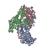 7qvxMC 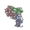 7qvyC 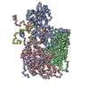 7qw9C M: atomic model generated by this map C: citing same article ( |
|---|---|
| Similar structure data |
-Links
| EMDB pages | EMDB (EBI/PDBe) / EMDataResource |
|---|---|
| Related items in Molecule of the Month |


-Map
| File | Download / File: emd_14183.map.gz / Format: CCP4 / Size: 125 MB / Type: IMAGE STORED AS FLOATING POINT NUMBER (4 BYTES) | ||||||||||||||||||||||||||||||||||||
|---|---|---|---|---|---|---|---|---|---|---|---|---|---|---|---|---|---|---|---|---|---|---|---|---|---|---|---|---|---|---|---|---|---|---|---|---|---|
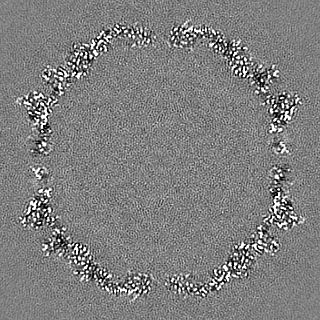
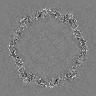
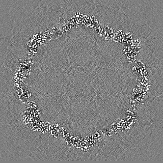
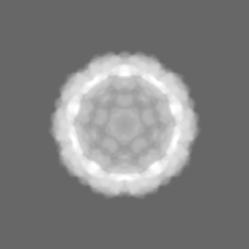
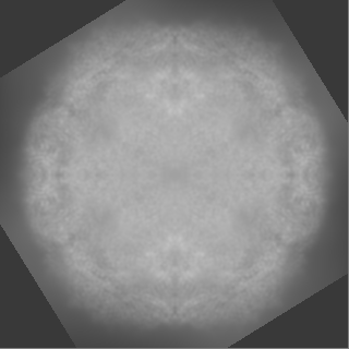
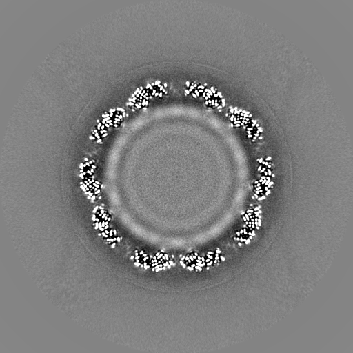
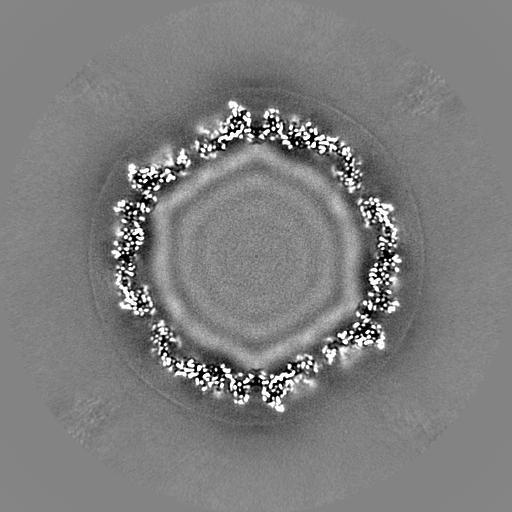
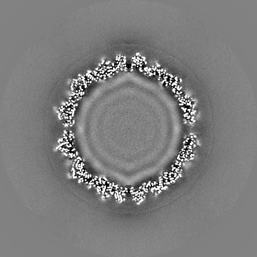
| Annotation | Sharpened map of CV-A6 altered particle at 2.5 A resolution | ||||||||||||||||||||||||||||||||||||
| Projections & slices | Image control
Images are generated by Spider. | ||||||||||||||||||||||||||||||||||||
| Voxel size | X=Y=Z: 1.063 Å | ||||||||||||||||||||||||||||||||||||
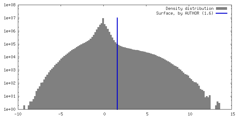
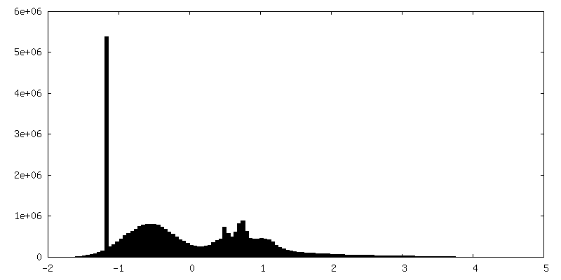
| Density |
| ||||||||||||||||||||||||||||||||||||
| Symmetry | Space group: 1 | ||||||||||||||||||||||||||||||||||||
| Details | EMDB XML:
|
 Z (Sec.)
 Y (Row.)
 X (Col.)


-Supplemental data
-Additional map: Mask used for calculation of the FSC curve by RELION postprocessing
| File | emd_14183_additional_1.map | ||||||||||||
|---|---|---|---|---|---|---|---|---|---|---|---|---|---|
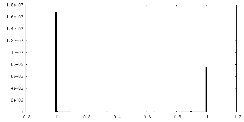
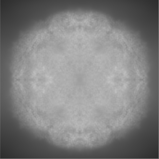
| Annotation | Mask used for calculation of the FSC curve by RELION postprocessing | ||||||||||||
| Projections & Slices |
| ||||||||||||
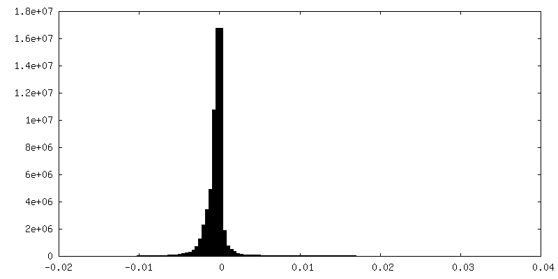
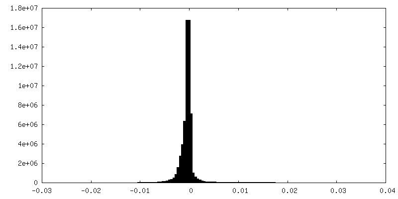
| Density Histograms |


-Additional map: Unsharpened map of CV-A6 altered particle at 2.7 A resolution
| File | emd_14183_additional_2.map | ||||||||||||
|---|---|---|---|---|---|---|---|---|---|---|---|---|---|
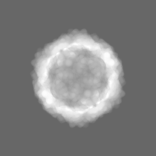
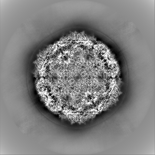
| Annotation | Unsharpened map of CV-A6 altered particle at 2.7 A resolution | ||||||||||||
| Projections & Slices |
| ||||||||||||
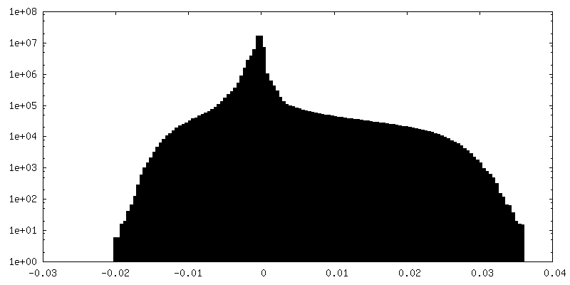
| Density Histograms |


-Half map: Ewald sphere corrected half map 1 used in RELION postprocessing
| File | emd_14183_half_map_1.map | ||||||||||||
|---|---|---|---|---|---|---|---|---|---|---|---|---|---|
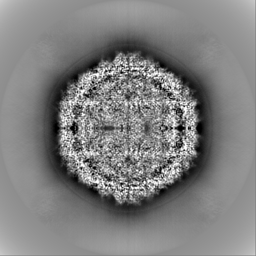
| Annotation | Ewald sphere corrected half map 1 used in RELION postprocessing | ||||||||||||
| Projections & Slices |
| ||||||||||||
| Density Histograms |


-Half map: Ewald sphere corrected half map 2 used in RELION postprocessing
| File | emd_14183_half_map_2.map | ||||||||||||
|---|---|---|---|---|---|---|---|---|---|---|---|---|---|
| Annotation | Ewald sphere corrected half map 2 used in RELION postprocessing | ||||||||||||
| Projections & Slices |
| ||||||||||||
| Density Histograms |


- Sample components
-Entire : Coxsackievirus A6
| Entire | Name: Coxsackievirus A6 |
|---|---|
| Components |
|
-Supramolecule #1: Coxsackievirus A6
| Supramolecule | Name: Coxsackievirus A6 / type: complex / ID: 1 / Parent: 0 / Macromolecule list: all / Details: expanded altered CV-A6 particle |
|---|---|
| Source (natural) | Organism: Coxsackievirus A6 / Strain: Gdula |
| Molecular weight | Theoretical: 8 MDa |
-Macromolecule #1: Capsid protein VP1
| Macromolecule | Name: Capsid protein VP1 / type: protein_or_peptide / ID: 1 Details: The first amino-acid of VP1 in the corresponding section of the GenBank polyprotein entry (AAR38844) (Asn) is not the actual first residue as the proteolytic cleavage of the viral ...Details: The first amino-acid of VP1 in the corresponding section of the GenBank polyprotein entry (AAR38844) (Asn) is not the actual first residue as the proteolytic cleavage of the viral polyprotein occurs at an alternative location, leaving the following residue Asp-2 as the first residue (D-1), and the Asn as the additional C-term residue described for VP3 (N-241), as observed in the virion. Number of copies: 1 / Enantiomer: LEVO |
|---|---|
| Source (natural) | Organism: Coxsackievirus A6 |
| Molecular weight | Theoretical: 33.530293 KDa |
| Sequence | String: DPISNAIENA VSTLADTTIS RVTAANTAAS SHSLGTGRVP ALQAAETGAS SNASDENLIE TRCVMNRNGV NEASVEHFYS RAGLVGVVE VKDSGTSQDG YTVWPIDVMG FVQQRRKLEL STYMRFDAEF TFVSNLNDST TPGMLLQYMY VPPGAPKPDG R KSYQWQTA ...String: DPISNAIENA VSTLADTTIS RVTAANTAAS SHSLGTGRVP ALQAAETGAS SNASDENLIE TRCVMNRNGV NEASVEHFYS RAGLVGVVE VKDSGTSQDG YTVWPIDVMG FVQQRRKLEL STYMRFDAEF TFVSNLNDST TPGMLLQYMY VPPGAPKPDG R KSYQWQTA TNPSIFAKLS DPPPQVSVPF MSPASAYQWF YDGYPTFGEH KQATNLQYGQ CPNNMMGHFA IRTVSESTTG KN VHVRVYM RIKHVRAWVP RPFRSQAYMV KNYPTYSQTI SNTAADRASI TTTDYEGGVP ANPQRTF UniProtKB: Genome polyprotein |
-Macromolecule #2: Capsid protein VP2
| Macromolecule | Name: Capsid protein VP2 / type: protein_or_peptide / ID: 2 / Number of copies: 1 / Enantiomer: LEVO |
|---|---|
| Source (natural) | Organism: Coxsackievirus A6 |
| Molecular weight | Theoretical: 28.006529 KDa |
| Sequence | String: SPSVEACGYS DRVAQLTVGN STITTQEAAN IVLSYGEWPG YCPSTDATAV DKPTRPDVSV NRFYTLSTKS WKTESTGWYW KFPDVLNDT GVFGQNAQFH YLYRSGFCMH VQCNASKFHQ GALLVVVIPE FVVAASSPAT KPNGQGLYPD FAHTNPGKEG Q VFRDPYVL ...String: SPSVEACGYS DRVAQLTVGN STITTQEAAN IVLSYGEWPG YCPSTDATAV DKPTRPDVSV NRFYTLSTKS WKTESTGWYW KFPDVLNDT GVFGQNAQFH YLYRSGFCMH VQCNASKFHQ GALLVVVIPE FVVAASSPAT KPNGQGLYPD FAHTNPGKEG Q VFRDPYVL DAGIPLSQAL VFPHQWINLR TNNCATIIMP YVNALPFDSA LNHSNFGLAV IPISPLKYCN GATTEVPITL TI APLNSEF SGLRQAIKQ UniProtKB: Genome polyprotein |
-Macromolecule #3: Capsid protein VP3
| Macromolecule | Name: Capsid protein VP3 / type: protein_or_peptide / ID: 3 Details: VP3 has an additional C-terminal residue (N-241) compared to GenBank entry AAR38844 due to alternative cleavage of the polyprotein as observed in the CV-A6 virion (see compound details for molecule 1 VP1). Number of copies: 1 / Enantiomer: LEVO |
|---|---|
| Source (natural) | Organism: Coxsackievirus A6 |
| Molecular weight | Theoretical: 26.489936 KDa |
| Sequence | String: GLPTELKPGT NQFLTTDDGT SPPILPGFEP TPLIHIPGEF TSLLDLCRIE TILEVNNTTG TTGVNRLLIP VRAQNNVDQL CASFQVDPG RNGPWQSTMV GQICRYYTQW SGSLKVTFMF TGSFMATGKM LIAYTPPGSA QPTTREAAML GTHIVWDFGL Q SSVTLVIP ...String: GLPTELKPGT NQFLTTDDGT SPPILPGFEP TPLIHIPGEF TSLLDLCRIE TILEVNNTTG TTGVNRLLIP VRAQNNVDQL CASFQVDPG RNGPWQSTMV GQICRYYTQW SGSLKVTFMF TGSFMATGKM LIAYTPPGSA QPTTREAAML GTHIVWDFGL Q SSVTLVIP WISNTHFRAV KTGGVYDYYA TGIVTIWYQT NFVVPPDTPS EANIIALGAA QENFTLKLCK DTDEIRQTAE YQ N UniProtKB: Genome polyprotein |
-Experimental details
-Structure determination
| Method | cryo EM |
|---|---|
 Processing Processing | single particle reconstruction |
| Aggregation state | particle |
-Sample preparation
| Concentration | 2.5 mg/mL | |||||||||||||||
|---|---|---|---|---|---|---|---|---|---|---|---|---|---|---|---|---|
| Buffer | pH: 7.4 Component:
Details: PBS, pH 7.4 | |||||||||||||||
| Grid | Model: Quantifoil R2/1 / Material: COPPER / Mesh: 300 / Support film - Material: CARBON / Support film - topology: HOLEY / Support film - Film thickness: 12 / Pretreatment - Type: GLOW DISCHARGE / Pretreatment - Time: 10 sec. | |||||||||||||||
| Vitrification | Cryogen name: ETHANE-PROPANE / Chamber humidity: 100 % / Chamber temperature: 283.15 K / Instrument: FEI VITROBOT MARK IV |
- Electron microscopy
| Microscope | FEI TITAN KRIOS |
|---|---|
| Image recording | Film or detector model: FEI FALCON II (4k x 4k) / Detector mode: INTEGRATING / Digitization - Dimensions - Width: 4096 pixel / Digitization - Dimensions - Height: 4096 pixel / Number grids imaged: 1 / Number real images: 9862 / Average exposure time: 1.0 sec. / Average electron dose: 45.0 e/Å2 |
| Electron beam | Acceleration voltage: 300 kV / Electron source:  FIELD EMISSION GUN FIELD EMISSION GUN |
| Electron optics | C2 aperture diameter: 70.0 µm / Calibrated defocus max: 4.3 µm / Calibrated defocus min: 0.3 µm / Illumination mode: FLOOD BEAM / Imaging mode: BRIGHT FIELD / Cs: 2.7 mm / Nominal defocus max: 3.0 µm / Nominal defocus min: 1.0 µm / Nominal magnification: 75000 |
| Sample stage | Specimen holder model: FEI TITAN KRIOS AUTOGRID HOLDER / Cooling holder cryogen: NITROGEN |
| Experimental equipment |  Model: Titan Krios / Image courtesy: FEI Company |
+Image processing

-Atomic model buiding 1
| Initial model | PDB ID: Chain - Source name: PDB / Chain - Initial model type: experimental model |
|---|---|
| Details | iterative cycles of building - real space refinement - reciprocal space refinement |
| Refinement | Space: RECIPROCAL / Protocol: RIGID BODY FIT / Overall B value: 29 / Target criteria: Correlation coefficient |
| Output model | PDB-7qvx: |

