+ Open data
- Basic information
| Entry | Database: PDB / ID: 7qvy | |||||||||
|---|---|---|---|---|---|---|---|---|---|---|
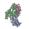
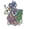
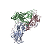
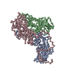
| Title | Cryo-EM structure of coxsackievirus A6 empty particle | |||||||||
 Components Components |
| |||||||||
 Keywords Keywords | VIRUS / enterovirus / coxsackievirus A6 / empty particle / capsid / cryo-EM | |||||||||
| Function / homology |  Function and homology information Function and homology informationsymbiont-mediated suppression of host cytoplasmic pattern recognition receptor signaling pathway via inhibition of MDA-5 activity / picornain 2A / symbiont-mediated suppression of host mRNA export from nucleus / symbiont genome entry into host cell via pore formation in plasma membrane / picornain 3C / T=pseudo3 icosahedral viral capsid / ribonucleoside triphosphate phosphatase activity / host cell cytoplasmic vesicle membrane / nucleoside-triphosphate phosphatase / channel activity ...symbiont-mediated suppression of host cytoplasmic pattern recognition receptor signaling pathway via inhibition of MDA-5 activity / picornain 2A / symbiont-mediated suppression of host mRNA export from nucleus / symbiont genome entry into host cell via pore formation in plasma membrane / picornain 3C / T=pseudo3 icosahedral viral capsid / ribonucleoside triphosphate phosphatase activity / host cell cytoplasmic vesicle membrane / nucleoside-triphosphate phosphatase / channel activity / monoatomic ion transmembrane transport / DNA replication / RNA helicase activity / endocytosis involved in viral entry into host cell / symbiont-mediated activation of host autophagy / RNA-directed RNA polymerase / cysteine-type endopeptidase activity / viral RNA genome replication / RNA-directed RNA polymerase activity / DNA-templated transcription / virion attachment to host cell / host cell nucleus / structural molecule activity / proteolysis / RNA binding / zinc ion binding / ATP binding / membrane Similarity search - Function | |||||||||
| Biological species |  Coxsackievirus A6 Coxsackievirus A6 | |||||||||
| Method | ELECTRON MICROSCOPY / single particle reconstruction / cryo EM / Resolution: 2.82 Å | |||||||||
 Authors Authors | Buttner, C.R. / Spurny, R. / Fuzik, T. / Plevka, P. | |||||||||
| Funding support |  Czech Republic, 2items Czech Republic, 2items
| |||||||||
 Citation Citation |  Journal: Commun Biol / Year: 2022 Journal: Commun Biol / Year: 2022Title: Cryo-electron microscopy and image classification reveal the existence and structure of the coxsackievirus A6 virion. Authors: Carina R Büttner / Radovan Spurný / Tibor Füzik / Pavel Plevka /  Abstract: Coxsackievirus A6 (CV-A6) has recently overtaken enterovirus A71 and CV-A16 as the primary causative agent of hand, foot, and mouth disease worldwide. Virions of CV-A6 were not identified in previous ...Coxsackievirus A6 (CV-A6) has recently overtaken enterovirus A71 and CV-A16 as the primary causative agent of hand, foot, and mouth disease worldwide. Virions of CV-A6 were not identified in previous structural studies, and it was speculated that the virus is unique among enteroviruses in using altered particles with expanded capsids to infect cells. In contrast, the virions of other enteroviruses are required for infection. Here we used cryo-electron microscopy (cryo-EM) to determine the structures of the CV-A6 virion, altered particle, and empty capsid. We show that the CV-A6 virion has features characteristic of virions of other enteroviruses, including a compact capsid, VP4 attached to the inner capsid surface, and fatty acid-like molecules occupying the hydrophobic pockets in VP1 subunits. Furthermore, we found that in a purified sample of CV-A6, the ratio of infectious units to virions is 1 to 500. Therefore, it is likely that virions of CV-A6 initiate infection, like those of other enteroviruses. Our results provide evidence that future vaccines against CV-A6 should target its virions instead of the antigenically distinct altered particles. Furthermore, the structure of the virion provides the basis for the rational development of capsid-binding inhibitors that block the genome release of CV-A6. | |||||||||
| History |
|
- Structure visualization
| Structure viewer | Molecule:  Molmil Molmil Jmol/JSmol Jmol/JSmol |
|---|
- Downloads & links
- Download
| PDBx/mmCIF format |  7qvy.cif.gz 7qvy.cif.gz | 139.5 KB | Display |  PDBx/mmCIF format PDBx/mmCIF format |
|---|---|---|---|---|
| PDB format |  pdb7qvy.ent.gz pdb7qvy.ent.gz | 106.8 KB | Display |  PDB format PDB format |
| PDBx/mmJSON format |  7qvy.json.gz 7qvy.json.gz | Tree view |  PDBx/mmJSON format PDBx/mmJSON format | |
| Others |  Other downloads Other downloads |
-Validation report
| Summary document |  7qvy_validation.pdf.gz 7qvy_validation.pdf.gz | 1.5 MB | Display |  wwPDB validaton report wwPDB validaton report |
|---|---|---|---|---|
| Full document |  7qvy_full_validation.pdf.gz 7qvy_full_validation.pdf.gz | 1.5 MB | Display | |
| Data in XML |  7qvy_validation.xml.gz 7qvy_validation.xml.gz | 41.8 KB | Display | |
| Data in CIF |  7qvy_validation.cif.gz 7qvy_validation.cif.gz | 59.9 KB | Display | |
| Arichive directory |  https://data.pdbj.org/pub/pdb/validation_reports/qv/7qvy https://data.pdbj.org/pub/pdb/validation_reports/qv/7qvy ftp://data.pdbj.org/pub/pdb/validation_reports/qv/7qvy ftp://data.pdbj.org/pub/pdb/validation_reports/qv/7qvy | HTTPS FTP |
-Related structure data
| Related structure data |  14184MC  7qvxC  7qw9C M: map data used to model this data C: citing same article ( |
|---|---|
| Similar structure data | Similarity search - Function & homology  F&H Search F&H Search |
- Links
Links
- Assembly
| Deposited unit | 
|
|---|---|
| 1 | x 60
|
- Components
| #1: Protein | Mass: 33530.293 Da / Num. of mol.: 1 / Source method: isolated from a natural source Details: The first amino-acid of VP1 in the corresponding section of the GenBank polyprotein entry (AAR38844) (Asn) is not the actual first residue as the proteolytic cleavage of the viral ...Details: The first amino-acid of VP1 in the corresponding section of the GenBank polyprotein entry (AAR38844) (Asn) is not the actual first residue as the proteolytic cleavage of the viral polyprotein occurs at an alternative location, leaving the following residue Asp-2 as the first residue (D-1), and the Asn as the extra C-term residue described for VP3 (N-241) as it was observed in the virion. Source: (natural)  Coxsackievirus A6 / References: UniProt: Q6JKS2 Coxsackievirus A6 / References: UniProt: Q6JKS2 |
|---|---|
| #2: Protein | Mass: 28006.529 Da / Num. of mol.: 1 / Source method: isolated from a natural source / Source: (natural)  Coxsackievirus A6 / References: UniProt: Q6JKS2 Coxsackievirus A6 / References: UniProt: Q6JKS2 |
| #3: Protein | Mass: 26489.936 Da / Num. of mol.: 1 / Source method: isolated from a natural source Details: VP3 has an additional C-terminal residue (N-241) compared to GenBank entry AAR38844 due to alternative cleavage of the polyprotein (see compound details for molecule 1 VP1). Source: (natural)  Coxsackievirus A6 / References: UniProt: Q6JKS2 Coxsackievirus A6 / References: UniProt: Q6JKS2 |
-Experimental details
-Experiment
| Experiment | Method: ELECTRON MICROSCOPY |
|---|---|
| EM experiment | Aggregation state: PARTICLE / 3D reconstruction method: single particle reconstruction |
- Sample preparation
| Component |
| |||||||||||||||||||||||||
|---|---|---|---|---|---|---|---|---|---|---|---|---|---|---|---|---|---|---|---|---|---|---|---|---|---|---|
| Molecular weight |
| |||||||||||||||||||||||||
| Source (natural) |
| |||||||||||||||||||||||||
| Details of virus | Empty: YES / Enveloped: NO / Isolate: STRAIN / Type: VIRION | |||||||||||||||||||||||||
| Natural host | Organism: Homo sapiens | |||||||||||||||||||||||||
| Virus shell | Name: CV-A6 empty particle / Diameter: 335 nm / Triangulation number (T number): 3 | |||||||||||||||||||||||||
| Buffer solution | pH: 7.4 / Details: PBS, pH 7.4 | |||||||||||||||||||||||||
| Buffer component |
| |||||||||||||||||||||||||
| Specimen | Conc.: 2.5 mg/ml / Embedding applied: NO / Shadowing applied: NO / Staining applied: NO / Vitrification applied: YES | |||||||||||||||||||||||||
| Specimen support | Grid material: COPPER / Grid mesh size: 300 divisions/in. / Grid type: Quantifoil R2/1 | |||||||||||||||||||||||||
| Vitrification | Instrument: FEI VITROBOT MARK IV / Cryogen name: ETHANE-PROPANE / Humidity: 100 % / Chamber temperature: 283.15 K |
- Electron microscopy imaging
| Experimental equipment |  Model: Titan Krios / Image courtesy: FEI Company |
|---|---|
| Microscopy | Model: FEI TITAN KRIOS |
| Electron gun | Electron source:  FIELD EMISSION GUN / Accelerating voltage: 300 kV / Illumination mode: FLOOD BEAM FIELD EMISSION GUN / Accelerating voltage: 300 kV / Illumination mode: FLOOD BEAM |
| Electron lens | Mode: BRIGHT FIELD / Nominal magnification: 75000 X / Nominal defocus max: 3000 nm / Nominal defocus min: 1000 nm / Calibrated defocus min: 500 nm / Calibrated defocus max: 3500 nm / Cs: 2.7 mm / C2 aperture diameter: 70 µm / Alignment procedure: COMA FREE |
| Specimen holder | Cryogen: NITROGEN / Specimen holder model: FEI TITAN KRIOS AUTOGRID HOLDER |
| Image recording | Average exposure time: 1 sec. / Electron dose: 45 e/Å2 / Detector mode: INTEGRATING / Film or detector model: FEI FALCON II (4k x 4k) / Num. of grids imaged: 1 / Num. of real images: 9862 |
| Image scans | Width: 4096 / Height: 4096 / Movie frames/image: 7 |
- Processing
| EM software |
| ||||||||||||||||||||||||||||||||||||||||||||||||||||||||||||
|---|---|---|---|---|---|---|---|---|---|---|---|---|---|---|---|---|---|---|---|---|---|---|---|---|---|---|---|---|---|---|---|---|---|---|---|---|---|---|---|---|---|---|---|---|---|---|---|---|---|---|---|---|---|---|---|---|---|---|---|---|---|
| CTF correction | Type: PHASE FLIPPING AND AMPLITUDE CORRECTION | ||||||||||||||||||||||||||||||||||||||||||||||||||||||||||||
| Particle selection | Num. of particles selected: 211991 | ||||||||||||||||||||||||||||||||||||||||||||||||||||||||||||
| Symmetry | Point symmetry: I (icosahedral) | ||||||||||||||||||||||||||||||||||||||||||||||||||||||||||||
| 3D reconstruction | Resolution: 2.82 Å / Resolution method: FSC 0.143 CUT-OFF / Num. of particles: 10613 / Algorithm: BACK PROJECTION / Num. of class averages: 1 / Symmetry type: POINT | ||||||||||||||||||||||||||||||||||||||||||||||||||||||||||||
| Atomic model building | B value: 37 / Protocol: RIGID BODY FIT / Space: RECIPROCAL / Target criteria: Correlation coefficient Details: iterative cycles of building - real space refinement - reciprocal space refinement | ||||||||||||||||||||||||||||||||||||||||||||||||||||||||||||
| Atomic model building | PDB-ID: 5XS4 Accession code: 5XS4 / Source name: PDB / Type: experimental model |
 Movie
Movie Controller


 PDBj
PDBj


