Movie
Movie Controller

[English] 日本語
 Yorodumi
- PDB-7pdz: Structure of capping protein bound to the barbed end of a cytopla... -
+ Open data

- Basic information
| Entry | Database: PDB / ID: 7pdz | |||||||||||||||||||||||||||||||||||||||||||||||||||||||||
|---|---|---|---|---|---|---|---|---|---|---|---|---|---|---|---|---|---|---|---|---|---|---|---|---|---|---|---|---|---|---|---|---|---|---|---|---|---|---|---|---|---|---|---|---|---|---|---|---|---|---|---|---|---|---|---|---|---|---|
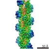
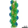
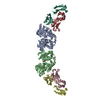
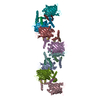
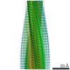
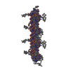
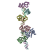
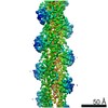
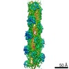
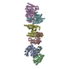
| Title | Structure of capping protein bound to the barbed end of a cytoplasmic actin filament | |||||||||||||||||||||||||||||||||||||||||||||||||||||||||
 Components Components |
| |||||||||||||||||||||||||||||||||||||||||||||||||||||||||
 Keywords Keywords | STRUCTURAL PROTEIN / Cytoskeleton / cell-shape remodelling / barbed end | |||||||||||||||||||||||||||||||||||||||||||||||||||||||||
| Function / homology |  Function and homology information Function and homology informationcytoskeletal calyx / Cell-extracellular matrix interactions / Formation of the canonical BAF (cBAF) complex / Formation of the polybromo-BAF (pBAF) complex / Formation of the embryonic stem cell BAF (esBAF) complex / Formation of the non-canonical BAF (ncBAF) complex / Formation of neuronal progenitor and neuronal BAF (npBAF and nBAF) / Adherens junctions interactions / Regulation of CDH1 Function / Formation of the dystrophin-glycoprotein complex (DGC) ...cytoskeletal calyx / Cell-extracellular matrix interactions / Formation of the canonical BAF (cBAF) complex / Formation of the polybromo-BAF (pBAF) complex / Formation of the embryonic stem cell BAF (esBAF) complex / Formation of the non-canonical BAF (ncBAF) complex / Formation of neuronal progenitor and neuronal BAF (npBAF and nBAF) / Adherens junctions interactions / Regulation of CDH1 Function / Formation of the dystrophin-glycoprotein complex (DGC) / Advanced glycosylation endproduct receptor signaling / B-WICH complex positively regulates rRNA expression / RHOF GTPase cycle / : / Gap junction degradation / Formation of annular gap junctions / RHOF GTPase cycle / EPHB-mediated forward signaling / negative regulation of filopodium assembly / MAP2K and MAPK activation / Regulation of actin dynamics for phagocytic cup formation / RHO GTPases activate IQGAPs / RHO GTPases Activate WASPs and WAVEs / COPI-independent Golgi-to-ER retrograde traffic / RHO GTPases Activate Formins / HSP90 chaperone cycle for steroid hormone receptors (SHR) in the presence of ligand / DNA Damage Recognition in GG-NER / COPI-mediated anterograde transport / UCH proteinases / F-actin capping protein complex / WASH complex / sperm head / VEGFA-VEGFR2 Pathway / Factors involved in megakaryocyte development and platelet production / cellular response to cytochalasin B / Clathrin-mediated endocytosis / regulation of transepithelial transport / MHC class II antigen presentation / morphogenesis of a polarized epithelium / cell junction assembly / structural constituent of postsynaptic actin cytoskeleton / protein localization to adherens junction / muscle cell development / barbed-end actin filament capping / actin polymerization or depolymerization / dense body / Tat protein binding / regulation of lamellipodium assembly / regulation of cell morphogenesis / postsynaptic actin cytoskeleton / adherens junction assembly / cell projection organization / apical protein localization / sperm head-tail coupling apparatus / lamellipodium assembly / tight junction / apical junction complex / negative regulation of microtubule polymerization / regulation of norepinephrine uptake / transporter regulator activity / NuA4 histone acetyltransferase complex / cortical cytoskeleton / establishment or maintenance of cell polarity / nitric-oxide synthase binding / brush border / intercalated disc / regulation of synaptic vesicle endocytosis / kinesin binding / regulation of protein localization to plasma membrane / positive regulation of double-strand break repair via homologous recombination / beta-tubulin binding / axonogenesis / cytoskeleton organization / calyx of Held / nitric-oxide synthase regulator activity / hippocampal mossy fiber to CA3 synapse / cell motility / actin filament / adherens junction / cell morphogenesis / Hydrolases; Acting on acid anhydrides; Acting on acid anhydrides to facilitate cellular and subcellular movement / Schaffer collateral - CA1 synapse / Z disc / neuron projection development / cytoplasmic ribonucleoprotein granule / cell-cell junction / actin filament binding / nucleosome / lamellipodium / actin cytoskeleton / actin cytoskeleton organization / actin binding / dendritic spine / cytoskeleton / regulation of cell cycle / postsynaptic density / ribonucleoprotein complex / axon / focal adhesion / neuronal cell body Similarity search - Function | |||||||||||||||||||||||||||||||||||||||||||||||||||||||||
| Biological species |  synthetic construct (others) | |||||||||||||||||||||||||||||||||||||||||||||||||||||||||
| Method | ELECTRON MICROSCOPY / single particle reconstruction / cryo EM / Resolution: 3.8 Å | |||||||||||||||||||||||||||||||||||||||||||||||||||||||||
 Authors Authors | Funk, J. / Merino, F. / Schacks, M. / Rottner, K. / Raunser, S. / Bieling, P. | |||||||||||||||||||||||||||||||||||||||||||||||||||||||||
| Funding support |  Germany, 1items Germany, 1items
| |||||||||||||||||||||||||||||||||||||||||||||||||||||||||
 Citation Citation | Journal: Nat Commun / Year: 2021 Title: A barbed end interference mechanism reveals how capping protein promotes nucleation in branched actin networks. Authors: Johanna Funk / Felipe Merino / Matthias Schaks / Klemens Rottner / Stefan Raunser / Peter Bieling / Abstract: Heterodimeric capping protein (CP/CapZ) is an essential factor for the assembly of branched actin networks, which push against cellular membranes to drive a large variety of cellular processes. Aside ...Heterodimeric capping protein (CP/CapZ) is an essential factor for the assembly of branched actin networks, which push against cellular membranes to drive a large variety of cellular processes. Aside from terminating filament growth, CP potentiates the nucleation of actin filaments by the Arp2/3 complex in branched actin networks through an unclear mechanism. Here, we combine structural biology with in vitro reconstitution to demonstrate that CP not only terminates filament elongation, but indirectly stimulates the activity of Arp2/3 activating nucleation promoting factors (NPFs) by preventing their association to filament barbed ends. Key to this function is one of CP's C-terminal "tentacle" extensions, which sterically masks the main interaction site of the terminal actin protomer. Deletion of the β tentacle only modestly impairs capping. However, in the context of a growing branched actin network, its removal potently inhibits nucleation promoting factors by tethering them to capped filament ends. End tethering of NPFs prevents their loading with actin monomers required for activation of the Arp2/3 complex and thus strongly inhibits branched network assembly both in cells and reconstituted motility assays. Our results mechanistically explain how CP couples two opposed processes-capping and nucleation-in branched actin network assembly. | |||||||||||||||||||||||||||||||||||||||||||||||||||||||||
| History |
|
- Structure visualization
| Movie |
Movie viewer |
|---|---|
| Structure viewer | Molecule: MolmilJmol/JSmol |
- Downloads & links
-Download
| PDBx/mmCIF format | 7pdz.cif.gz | 488.8 KB | Display | PDBx/mmCIF format |
|---|---|---|---|---|
| PDB format | pdb7pdz.ent.gz | 404.6 KB | Display | PDB format |
| PDBx/mmJSON format | 7pdz.json.gz | Tree view | PDBx/mmJSON format | |
| Others |  Other downloads Other downloads |
-Validation report
| Arichive directory | https://data.pdbj.org/pub/pdb/validation_reports/pd/7pdzftp://data.pdbj.org/pub/pdb/validation_reports/pd/7pdz | HTTPS FTP |
|---|
-Related structure data
| Related structure data | 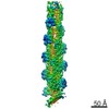 13343MC M: map data used to model this data C: citing same article ( |
|---|---|
| Similar structure data |
-Links
 PDBj
PDBj


- Assembly
| Deposited unit | 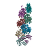
|
|---|---|
| 1 |
|
-Components
-Protein , 3 types, 8 molecules EFIJKLNO
| #1: Protein | Mass: 30669.768 Da / Num. of mol.: 1 Source method: isolated from a genetically manipulated source Source: (gene. exp.)  |
|---|---|
| #2: Protein | Mass: 32980.703 Da / Num. of mol.: 1 Source method: isolated from a genetically manipulated source Source: (gene. exp.) |
| #3: Protein | Mass: 41795.680 Da / Num. of mol.: 6 / Source method: isolated from a natural source / Source: (natural) |
-Protein/peptide , 1 types, 5 molecules QRSTP
| #4: Protein/peptide | 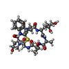  Type: Cyclic peptide / Class: Toxin / Mass: 808.899 Da / Num. of mol.: 5 / Source method: obtained synthetically / Source: (synth.) synthetic construct (others) / References: Phalloidin Type: Cyclic peptide / Class: Toxin / Mass: 808.899 Da / Num. of mol.: 5 / Source method: obtained synthetically / Source: (synth.) synthetic construct (others) / References: Phalloidin |
|---|
-Non-polymers , 3 types, 16 molecules 


| #5: Chemical | ChemComp-ADP /  Mass: 427.201 Da / Num. of mol.: 6 / Source method: obtained synthetically / Formula: C10H15N5O10P2 / Feature type: SUBJECT OF INVESTIGATION / Comment: ADP, energy-carrying molecule*YM Mass: 427.201 Da / Num. of mol.: 6 / Source method: obtained synthetically / Formula: C10H15N5O10P2 / Feature type: SUBJECT OF INVESTIGATION / Comment: ADP, energy-carrying molecule*YM#6: Chemical | ChemComp-MG /  Mass: 24.305 Da / Num. of mol.: 6 / Source method: obtained synthetically / Formula: Mg / Feature type: SUBJECT OF INVESTIGATION Mass: 24.305 Da / Num. of mol.: 6 / Source method: obtained synthetically / Formula: Mg / Feature type: SUBJECT OF INVESTIGATION#7: Chemical | ChemComp-PO4 /  Mass: 94.971 Da / Num. of mol.: 4 / Source method: obtained synthetically / Formula: PO4 / Feature type: SUBJECT OF INVESTIGATION Mass: 94.971 Da / Num. of mol.: 4 / Source method: obtained synthetically / Formula: PO4 / Feature type: SUBJECT OF INVESTIGATION |
|---|
-Details
| Has ligand of interest | Y |
|---|---|
| Has protein modification | Y |
-Experimental details
-Experiment
| Experiment | Method: ELECTRON MICROSCOPY |
|---|---|
| EM experiment | Aggregation state: PARTICLE / 3D reconstruction method: single particle reconstruction |
- Sample preparation
| Component | Name: Complex between capping protein and the barbed end of cytoplasmic actin filaments Type: COMPLEX / Entity ID: #1-#4 / Source: MULTIPLE SOURCES | |||||||||||||||||||||||||
|---|---|---|---|---|---|---|---|---|---|---|---|---|---|---|---|---|---|---|---|---|---|---|---|---|---|---|
| Molecular weight | Experimental value: NO | |||||||||||||||||||||||||
| Buffer solution | pH: 7 / Details: KMEI buffer | |||||||||||||||||||||||||
| Buffer component |
| |||||||||||||||||||||||||
| Specimen | Embedding applied: NO / Shadowing applied: NO / Staining applied: NO / Vitrification applied: YES | |||||||||||||||||||||||||
| Specimen support | Details: Mild discharging with 5 mA current / Grid material: COPPER / Grid type: Quantifoil R1.2/1.3 | |||||||||||||||||||||||||
| Vitrification | Instrument: FEI VITROBOT MARK IV / Cryogen name: ETHANE / Humidity: 100 % / Chamber temperature: 286 K |
- Electron microscopy imaging
| Experimental equipment |  Model: Talos Arctica / Image courtesy: FEI Company |
|---|---|
| Microscopy | Model: FEI TALOS ARCTICA |
| Electron gun | Electron source:  FIELD EMISSION GUN / Accelerating voltage: 200 kV / Illumination mode: FLOOD BEAM FIELD EMISSION GUN / Accelerating voltage: 200 kV / Illumination mode: FLOOD BEAM |
| Electron lens | Mode: BRIGHT FIELD / Cs: 2.7 mm / Alignment procedure: COMA FREE |
| Specimen holder | Cryogen: NITROGEN |
| Image recording | Average exposure time: 3 sec. / Electron dose: 60 e/Å2 / Detector mode: INTEGRATING / Film or detector model: FEI FALCON III (4k x 4k) / Num. of grids imaged: 2 / Num. of real images: 4204 |
- Processing
| EM software |
| ||||||||||||||||||||||||||||||||||||||||
|---|---|---|---|---|---|---|---|---|---|---|---|---|---|---|---|---|---|---|---|---|---|---|---|---|---|---|---|---|---|---|---|---|---|---|---|---|---|---|---|---|---|
| CTF correction | Type: PHASE FLIPPING AND AMPLITUDE CORRECTION | ||||||||||||||||||||||||||||||||||||||||
| Particle selection | Num. of particles selected: 570724 | ||||||||||||||||||||||||||||||||||||||||
| 3D reconstruction | Resolution: 3.8 Å / Resolution method: FSC 0.143 CUT-OFF / Num. of particles: 60206 / Algorithm: BACK PROJECTION / Num. of class averages: 1 / Symmetry type: POINT | ||||||||||||||||||||||||||||||||||||||||
| Atomic model building | B value: 40 / Protocol: FLEXIBLE FIT / Space: REAL | ||||||||||||||||||||||||||||||||||||||||
| Atomic model building | PDB-ID: 5ADX Accession code: 5ADX / Source name: PDB / Type: experimental model |