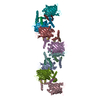
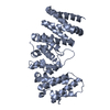
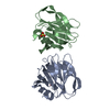
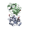
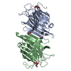
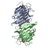
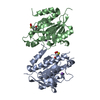
- PDB-7bwn: Crystal Structure of a Designed Protein Heterocatenane -
+
Open data
ID or keywords:
Loading...
-
Basic information
Entry
Database: PDB / ID: 7bwn
Title
Crystal Structure of a Designed Protein Heterocatenane
Components
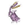
Cellular tumor antigen p53
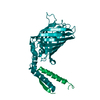
Chimera of Green fluorescent protein and p53dim
Keywords
RECOMBINATION / Heterocatenane
Function / homology
Function and homology information
negative regulation of helicase activity / Loss of function of TP53 in cancer due to loss of tetramerization ability / Regulation of TP53 Expression / signal transduction by p53 class mediator / negative regulation of G1 to G0 transition / negative regulation of glucose catabolic process to lactate via pyruvate / Transcriptional activation of cell cycle inhibitor p21 / regulation of intrinsic apoptotic signaling pathway by p53 class mediator / negative regulation of pentose-phosphate shunt / Activation of NOXA and translocation to mitochondria ...negative regulation of helicase activity / Loss of function of TP53 in cancer due to loss of tetramerization ability / Regulation of TP53 Expression / signal transduction by p53 class mediator / negative regulation of G1 to G0 transition / negative regulation of glucose catabolic process to lactate via pyruvate / Transcriptional activation of cell cycle inhibitor p21 / regulation of intrinsic apoptotic signaling pathway by p53 class mediator / negative regulation of pentose-phosphate shunt / Activation of NOXA and translocation to mitochondria / ATP-dependent DNA/DNA annealing activity / regulation of cell cycle G2/M phase transition / oligodendrocyte apoptotic process / negative regulation of miRNA processing / intrinsic apoptotic signaling pathway in response to hypoxia / oxidative stress-induced premature senescence / regulation of tissue remodeling / positive regulation of thymocyte apoptotic process / positive regulation of mitochondrial membrane permeability / germ cell nucleus / regulation of fibroblast apoptotic process / bone marrow development / cellular response to actinomycin D / circadian behavior / regulation of mitochondrial membrane permeability involved in apoptotic process / histone deacetylase regulator activity / positive regulation of programmed necrotic cell death / : / RUNX3 regulates CDKN1A transcription / T cell proliferation involved in immune response / TP53 Regulates Transcription of Death Receptors and Ligands / Activation of PUMA and translocation to mitochondria / TP53 regulates transcription of additional cell cycle genes whose exact role in the p53 pathway remain uncertain / mRNA transcription / negative regulation of glial cell proliferation / negative regulation of neuroblast proliferation / regulation of DNA damage response, signal transduction by p53 class mediator / Regulation of TP53 Activity through Association with Co-factors / Formation of Senescence-Associated Heterochromatin Foci (SAHF) / mitochondrial DNA repair / T cell lineage commitment / thymocyte apoptotic process / ER overload response / TP53 Regulates Transcription of Caspase Activators and Caspases / cardiac septum morphogenesis / necroptotic process / B cell lineage commitment / entrainment of circadian clock by photoperiod / negative regulation of DNA replication / Zygotic genome activation (ZGA) / negative regulation of mitophagy / TP53 Regulates Transcription of Genes Involved in Cytochrome C Release / PI5P Regulates TP53 Acetylation / positive regulation of release of cytochrome c from mitochondria / neuroblast proliferation / Association of TriC/CCT with target proteins during biosynthesis / negative regulation of telomere maintenance via telomerase / SUMOylation of transcription factors / TP53 regulates transcription of several additional cell death genes whose specific roles in p53-dependent apoptosis remain uncertain / rRNA transcription / negative regulation of reactive oxygen species metabolic process / intrinsic apoptotic signaling pathway by p53 class mediator / TFIID-class transcription factor complex binding / Transcriptional Regulation by VENTX / cellular response to UV-C / replicative senescence / viral process / intrinsic apoptotic signaling pathway in response to endoplasmic reticulum stress / hematopoietic stem cell differentiation / embryonic organ development / intrinsic apoptotic signaling pathway in response to DNA damage by p53 class mediator / Pyroptosis / positive regulation of RNA polymerase II transcription preinitiation complex assembly / chromosome organization / general transcription initiation factor binding / positive regulation of execution phase of apoptosis / type II interferon-mediated signaling pathway / hematopoietic progenitor cell differentiation / negative regulation of stem cell proliferation / TP53 Regulates Transcription of Genes Involved in G1 Cell Cycle Arrest / response to X-ray / somitogenesis / negative regulation of fibroblast proliferation / core promoter sequence-specific DNA binding / glial cell proliferation / cis-regulatory region sequence-specific DNA binding / cellular response to glucose starvation / mitophagy / Regulation of TP53 Activity through Acetylation / negative regulation of proteolysis / mitotic G1 DNA damage checkpoint signaling / positive regulation of intrinsic apoptotic signaling pathway / response to salt stress / cardiac muscle cell apoptotic process / transcription repressor complex / stem cell proliferation / gastrulation / 14-3-3 protein binding / positive regulation of cardiac muscle cell apoptotic process / transforming growth factor beta receptor signaling pathway Similarity search - Function
p53, subunit A / p53-like tetramerisation domain / Cellular tumor antigen p53, transactivation domain 2 / Transactivation domain 2 / p53 transactivation domain / P53 transactivation motif / : / p53 family signature. / p53, tetramerisation domain / P53 tetramerisation motif ...p53, subunit A / p53-like tetramerisation domain / Cellular tumor antigen p53, transactivation domain 2 / Transactivation domain 2 / p53 transactivation domain / P53 transactivation motif / : / p53 family signature. / p53, tetramerisation domain / P53 tetramerisation motif / p53, DNA-binding domain / P53 DNA-binding domain / p53 tumour suppressor family / p53-like tetramerisation domain superfamily / p53/RUNT-type transcription factor, DNA-binding domain superfamily / Green Fluorescent Protein / Green fluorescent protein / p53-like transcription factor, DNA-binding / Green fluorescent protein, GFP / Green fluorescent protein-related / Green fluorescent protein / Green fluorescent protein / Few Secondary Structures / Irregular / Beta Barrel / Mainly Beta Similarity search - Domain/homology
F: Chimera of Green fluorescent protein and p53dim L: Cellular tumor antigen p53 A: Chimera of Green fluorescent protein and p53dim B: Cellular tumor antigen p53 C: Chimera of Green fluorescent protein and p53dim D: Cellular tumor antigen p53 E: Chimera of Green fluorescent protein and p53dim G: Cellular tumor antigen p53 H: Chimera of Green fluorescent protein and p53dim I: Cellular tumor antigen p53 J: Chimera of Green fluorescent protein and p53dim K: Cellular tumor antigen p53 M: Chimera of Green fluorescent protein and p53dim N: Cellular tumor antigen p53 O: Chimera of Green fluorescent protein and p53dim P: Cellular tumor antigen p53
 Movie
Movie Controller


 Open data
 Basic information
 Components
 Keywords
 Function and homology information
 Aequorea victoria (jellyfish)
 Homo sapiens (human)
 X-RAY DIFFRACTION /
  Authors
 China, 3items
  Citation
 Structure visualization
 Downloads & links
 Other downloads
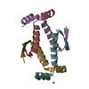


 PDBj
PDBj


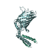
 Assembly