 Movie
Movie Controller

[English] 日本語
 Yorodumi
- PDB-6lsp: Crystal structure of a dimeric Piptidyl t-RNA hydrolase from Acin... -
+ Open data

- Basic information
| Entry | Database: PDB / ID: 6lsp | ||||||
|---|---|---|---|---|---|---|---|
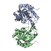
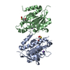
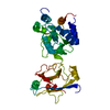
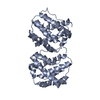
| Title | Crystal structure of a dimeric Piptidyl t-RNA hydrolase from Acinetobacter baumannii at 1.50 A resolution reveals an inhibited form. | ||||||
 Components Components | Peptidyl-tRNA hydrolase | ||||||
 Keywords Keywords | HYDROLASE | ||||||
| Function / homology |  Function and homology information Function and homology informationpeptidyl-tRNA hydrolase / peptidyl-tRNA hydrolase activity / protein quality control for misfolded or incompletely synthesized proteins / rescue of stalled cytosolic ribosome / tRNA binding / cytoplasm Similarity search - Function | ||||||
| Biological species |  Acinetobacter baumannii (bacteria) Acinetobacter baumannii (bacteria) | ||||||
| Method |  X-RAY DIFFRACTION / SYNCHROTRON / MOLECULAR REPLACEMENT / Resolution: 1.5 Å X-RAY DIFFRACTION / SYNCHROTRON / MOLECULAR REPLACEMENT / Resolution: 1.5 Å | ||||||
 Authors Authors | Viswanathan, V. / Ahmad, M.I. / Sharma, P. / Sharma, S. / Singh, T.P. | ||||||
| Funding support |  India, 1items India, 1items
| ||||||
 Citation Citation | Journal: To Be Published Title: Crystal structure of a dimeric Piptidyl t-RNA hydrolase from Acinetobacter baumannii at 1.50 A resolution reveals on inhibited form. Authors: Viswanathan, V. / Ahmad, M.I. / Sharma, P. / Sharma, S. / Singh, T.P. | ||||||
| History |
|
- Structure visualization
| Structure viewer | Molecule: MolmilJmol/JSmol |
|---|
- Downloads & links
-Download
| PDBx/mmCIF format | 6lsp.cif.gz | 123.6 KB | Display | PDBx/mmCIF format |
|---|---|---|---|---|
| PDB format | pdb6lsp.ent.gz | 75.7 KB | Display | PDB format |
| PDBx/mmJSON format | 6lsp.json.gz | Tree view | PDBx/mmJSON format | |
| Others |  Other downloads Other downloads |
-Validation report
| Arichive directory | https://data.pdbj.org/pub/pdb/validation_reports/ls/6lspftp://data.pdbj.org/pub/pdb/validation_reports/ls/6lsp | HTTPS FTP |
|---|
-Related structure data
| Related structure data | 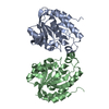 5yn4S S: Starting model for refinement |
|---|---|
| Similar structure data |


-Links
 PDBj
PDBj- Assembly
| Deposited unit | 
| ||||||||||||
|---|---|---|---|---|---|---|---|---|---|---|---|---|---|
| 1 |
| ||||||||||||
| Unit cell |
|
-Components
| #1: Protein | Mass: 20967.957 Da / Num. of mol.: 2 Source method: isolated from a genetically manipulated source Source: (gene. exp.) Acinetobacter baumannii (strain ATCC 19606 / DSM 30007 / CIP 70.34 / JCM 6841 / NBRC 109757 / NCIMB 12457 / NCTC 12156 / 81) (bacteria)Strain: ATCC 19606 / DSM 30007 / CIP 70.34 / JCM 6841 / NBRC 109757 / NCIMB 12457 / NCTC 12156 / 81 Gene: pth, BIT33_16330, HMPREF0010_01329 / Production host: #2: Water | ChemComp-HOH / |  Mass: 18.015 Da / Num. of mol.: 622 / Source method: isolated from a natural source / Formula: H2O Mass: 18.015 Da / Num. of mol.: 622 / Source method: isolated from a natural source / Formula: H2O |
|---|
-Experimental details
-Experiment
| Experiment | Method: X-RAY DIFFRACTION / Number of used crystals: 1 |
|---|
- Sample preparation
| Crystal | Density Matthews: 2.51 Å3/Da / Density % sol: 51.04 % |
|---|---|
| Crystal grow | Temperature: 293 K / Method: vapor diffusion, hanging drop / pH: 7.5 / Details: 20% PEG 1500, 0.1M HEPES, pH 7.5, 22% Glycerol |
-Data collection
| Diffraction | Mean temperature: 100 K / Serial crystal experiment: N |
|---|---|
| Diffraction source | Source: SYNCHROTRON / Site: RRCAT INDUS-2 / Beamline: PX-BL21 / Wavelength: 0.97 Å |
| Detector | Type: MARMOSAIC 225 mm CCD / Detector: CCD / Date: Dec 4, 2019 |
| Radiation | Protocol: SINGLE WAVELENGTH / Monochromatic (M) / Laue (L): M / Scattering type: x-ray |
| Radiation wavelength | Wavelength: 0.97 Å / Relative weight: 1 |
| Reflection | Resolution: 1.5→38.56 Å / Num. obs: 68761 / % possible obs: 99.97 % / Redundancy: 6.5 % / Biso Wilson estimate: 10.37 Å2 / CC1/2: 0.99 / Rmerge(I) obs: 0.1 / Rpim(I) all: 0.042 / Rrim(I) all: 0.108 / Net I/σ(I): 11.2 |
| Reflection shell | Resolution: 1.5→1.53 Å / Redundancy: 6.4 % / Rmerge(I) obs: 0.53 / Mean I/σ(I) obs: 3.4 / Num. unique obs: 4972 / CC1/2: 0.86 / Rpim(I) all: 0.22 / Rrim(I) all: 0.58 / % possible all: 99.92 |
- Processing
| Software |
| |||||||||||||||||||||||||||||||||||||||||||||||||||||||||||||||||||||||||||||
|---|---|---|---|---|---|---|---|---|---|---|---|---|---|---|---|---|---|---|---|---|---|---|---|---|---|---|---|---|---|---|---|---|---|---|---|---|---|---|---|---|---|---|---|---|---|---|---|---|---|---|---|---|---|---|---|---|---|---|---|---|---|---|---|---|---|---|---|---|---|---|---|---|---|---|---|---|---|---|
| Refinement | Method to determine structure: MOLECULAR REPLACEMENT Starting model: 5YN4 Resolution: 1.5→33.3 Å / SU ML: 0.1213 / Cross valid method: FREE R-VALUE / σ(F): 1.35 / Phase error: 17.9696
| |||||||||||||||||||||||||||||||||||||||||||||||||||||||||||||||||||||||||||||
| Solvent computation | Shrinkage radii: 0.9 Å / VDW probe radii: 1.11 Å | |||||||||||||||||||||||||||||||||||||||||||||||||||||||||||||||||||||||||||||
| Displacement parameters | Biso mean: 15.2 Å2 | |||||||||||||||||||||||||||||||||||||||||||||||||||||||||||||||||||||||||||||
| Refinement step | Cycle: LAST / Resolution: 1.5→33.3 Å
| |||||||||||||||||||||||||||||||||||||||||||||||||||||||||||||||||||||||||||||
| Refine LS restraints |
| |||||||||||||||||||||||||||||||||||||||||||||||||||||||||||||||||||||||||||||
| LS refinement shell |
|
