- PDB-4s0p: Crystal Structure of the Autoinhibited Dimer of Pro-apoptotic BAX (II) -
+
Open data
ID or keywords:
Loading...
-
Basic information
Entry
Database: PDB / ID: 4s0p
Title
Crystal Structure of the Autoinhibited Dimer of Pro-apoptotic BAX (II)
Components
Apoptosis regulator BAX
Keywords
APOPTOSIS / BCL-2 FAMILY PROTEIN / APOPTOSIS REGULATOR / AUTOINHIBITED DIMER
Function / homology
Function and homology information
release of matrix enzymes from mitochondria / BAX complex / B cell receptor apoptotic signaling pathway / Activation, translocation and oligomerization of BAX / B cell apoptotic process / positive regulation of apoptotic DNA fragmentation / NTRK3 as a dependence receptor / BAK complex / mitochondrial permeability transition pore complex / Release of apoptotic factors from the mitochondria ...release of matrix enzymes from mitochondria / BAX complex / B cell receptor apoptotic signaling pathway / Activation, translocation and oligomerization of BAX / B cell apoptotic process / positive regulation of apoptotic DNA fragmentation / NTRK3 as a dependence receptor / BAK complex / mitochondrial permeability transition pore complex / Release of apoptotic factors from the mitochondria / mitochondrial fragmentation involved in apoptotic process / Transcriptional regulation by RUNX2 / establishment or maintenance of transmembrane electrochemical gradient / endoplasmic reticulum calcium ion homeostasis / apoptotic mitochondrial changes / mitochondrial fusion / execution phase of apoptosis / Bcl-2 family protein complex / pore complex / extrinsic apoptotic signaling pathway via death domain receptors / positive regulation of IRE1-mediated unfolded protein response / TP53 Regulates Transcription of Genes Involved in Cytochrome C Release / positive regulation of release of cytochrome c from mitochondria / negative regulation of mitochondrial membrane potential / intrinsic apoptotic signaling pathway in response to endoplasmic reticulum stress / extrinsic apoptotic signaling pathway in absence of ligand / cellular response to unfolded protein / positive regulation of intrinsic apoptotic signaling pathway / BH3 domain binding / Pyroptosis / negative regulation of protein binding / extrinsic apoptotic signaling pathway / release of cytochrome c from mitochondria / intrinsic apoptotic signaling pathway / supramolecular fiber organization / TP53 Regulates Transcription of Genes Involved in G2 Cell Cycle Arrest / apoptotic signaling pathway / regulation of mitochondrial membrane potential / Hsp70 protein binding / intrinsic apoptotic signaling pathway in response to DNA damage / cellular response to virus / positive regulation of protein-containing complex assembly / response to toxic substance / positive regulation of neuron apoptotic process / nuclear envelope / channel activity / regulation of apoptotic process / mitochondrial outer membrane / positive regulation of apoptotic process / apoptotic process / protein heterodimerization activity / lipid binding / negative regulation of apoptotic process / endoplasmic reticulum membrane / endoplasmic reticulum / protein homodimerization activity / mitochondrion / extracellular exosome / membrane / identical protein binding / nucleus / cytosol / cytoplasm Similarity search - Function
Blc2-like / Apoptosis Regulator Bcl-x / Apoptosis regulator, Bcl-2, BH3 motif, conserved site / Apoptosis regulator, Bcl-2 family BH3 motif signature. / Apoptosis regulator, Bcl-2, BH1 motif, conserved site / Apoptosis regulator, Bcl-2 family BH1 motif signature. / Apoptosis regulator, Bcl-2, BH2 motif, conserved site / Apoptosis regulator, Bcl-2 family BH2 motif signature. / Bcl-2 family / BCL (B-Cell lymphoma); contains BH1, BH2 regions ...Blc2-like / Apoptosis Regulator Bcl-x / Apoptosis regulator, Bcl-2, BH3 motif, conserved site / Apoptosis regulator, Bcl-2 family BH3 motif signature. / Apoptosis regulator, Bcl-2, BH1 motif, conserved site / Apoptosis regulator, Bcl-2 family BH1 motif signature. / Apoptosis regulator, Bcl-2, BH2 motif, conserved site / Apoptosis regulator, Bcl-2 family BH2 motif signature. / Bcl-2 family / BCL (B-Cell lymphoma); contains BH1, BH2 regions / Bcl2-like / Bcl-2, Bcl-2 homology region 1-3 / Apoptosis regulator proteins, Bcl-2 family / BCL2-like apoptosis inhibitors family profile. / Bcl-2-like superfamily / Orthogonal Bundle / Mainly Alpha Similarity search - Domain/homology
In the structure databanks used in Yorodumi, some data are registered as the other names, "COVID-19 virus" and "2019-nCoV". Here are the details of the virus and the list of structure data.
Jan 31, 2019. EMDB accession codes are about to change! (news from PDBe EMDB page)
EMDB accession codes are about to change! (news from PDBe EMDB page)
The allocation of 4 digits for EMDB accession codes will soon come to an end. Whilst these codes will remain in use, new EMDB accession codes will include an additional digit and will expand incrementally as the available range of codes is exhausted. The current 4-digit format prefixed with “EMD-” (i.e. EMD-XXXX) will advance to a 5-digit format (i.e. EMD-XXXXX), and so on. It is currently estimated that the 4-digit codes will be depleted around Spring 2019, at which point the 5-digit format will come into force.
The EM Navigator/Yorodumi systems omit the EMD- prefix.
Related info.:Q: What is EMD? / ID/Accession-code notation in Yorodumi/EM Navigator
Yorodumi is a browser for structure data from EMDB, PDB, SASBDB, etc.
This page is also the successor to EM Navigator detail page, and also detail information page/front-end page for Omokage search.
The word "yorodu" (or yorozu) is an old Japanese word meaning "ten thousand". "mi" (miru) is to see.
Related info.:EMDB / PDB / SASBDB / Comparison of 3 databanks / Yorodumi Search / Aug 31, 2016. New EM Navigator & Yorodumi / Yorodumi Papers / Jmol/JSmol / Function and homology information / Changes in new EM Navigator and Yorodumi
 Movie
Movie Controller

 Yorodumi
 Open data
 Basic information
 Components
 Keywords
 Function and homology information
 Homo sapiens (human)
 X-RAY DIFFRACTION /
  Authors
 Citation
 Structure visualization
 Downloads & links
 Other downloads

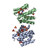
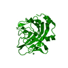
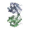
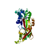


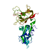
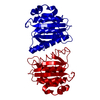
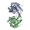
 PDBj
PDBj







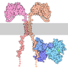
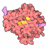

 Assembly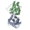
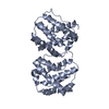


 Escherichia coli (E. coli) / References: UniProt: Q07812
 Sample preparation
 / Beamline: 23-ID-D / Wavelength: 1 Å
 Processing