 Movie
Movie Controller

[English] 日本語
 Yorodumi
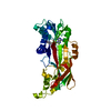
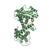
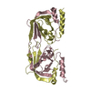
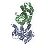
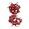
- PDB-2v24: Structure of the human SPRY domain-containing SOCS box protein SSB-4 -
+ Open data

- Basic information
| Entry | Database: PDB / ID: 2v24 | ||||||
|---|---|---|---|---|---|---|---|
| Title | Structure of the human SPRY domain-containing SOCS box protein SSB-4 | ||||||
 Components Components | SPRY DOMAIN-CONTAINING SOCS BOX PROTEIN 4 | ||||||
 Keywords Keywords | PROTEIN BINDING / PROTEIN-BINDING | ||||||
| Function / homology |  Function and homology information Function and homology informationpositive regulation of protein polyubiquitination / SCF ubiquitin ligase complex / ubiquitin-like ligase-substrate adaptor activity / regulation of circadian rhythm / rhythmic process / Antigen processing: Ubiquitination & Proteasome degradation / Neddylation / ubiquitin-dependent protein catabolic process / proteasome-mediated ubiquitin-dependent protein catabolic process / intracellular signal transduction ...positive regulation of protein polyubiquitination / SCF ubiquitin ligase complex / ubiquitin-like ligase-substrate adaptor activity / regulation of circadian rhythm / rhythmic process / Antigen processing: Ubiquitination & Proteasome degradation / Neddylation / ubiquitin-dependent protein catabolic process / proteasome-mediated ubiquitin-dependent protein catabolic process / intracellular signal transduction / protein ubiquitination / cytosol Similarity search - Function | ||||||
| Biological species |  HOMO SAPIENS (human) HOMO SAPIENS (human) | ||||||
| Method |  X-RAY DIFFRACTION / SYNCHROTRON / MOLECULAR REPLACEMENT / Resolution: 2.2 Å X-RAY DIFFRACTION / SYNCHROTRON / MOLECULAR REPLACEMENT / Resolution: 2.2 Å | ||||||
 Authors Authors | Uppenberg, J. / Bullock, A. / Keates, T. / Savitsky, P. / Pike, A.C.W. / Ugochukwu, E. / Bunkoczi, G. / von Delft, F. / Weigelt, J. / Arrowsmith, C.H. ...Uppenberg, J. / Bullock, A. / Keates, T. / Savitsky, P. / Pike, A.C.W. / Ugochukwu, E. / Bunkoczi, G. / von Delft, F. / Weigelt, J. / Arrowsmith, C.H. / Edwards, A. / Sundstrom, M. / Knapp, S. | ||||||
 Citation Citation | Journal: J.Mol.Biol. / Year: 2010 Title: Structural Basis for Par-4 Recognition by the Spry Domain-and Socs Box-Containing Proteins Spsb1, Spsb2, and Spsb4. Authors: Filippakopoulos, P. / Low, A. / Sharpe, T.D. / Uppenberg, J. / Yao, S. / Kuang, Z. / Savitsky, P. / Lewis, R.S. / Nicholson, S.E. / Norton, R.S. / Bullock, A. | ||||||
| History |
| ||||||
| Remark 700 | SHEET THE SHEET STRUCTURE OF THIS MOLECULE IS BIFURCATED. IN ORDER TO REPRESENT THIS FEATURE IN ... SHEET THE SHEET STRUCTURE OF THIS MOLECULE IS BIFURCATED. IN ORDER TO REPRESENT THIS FEATURE IN THE SHEET RECORDS BELOW, TWO SHEETS ARE DEFINED. |
- Structure visualization
| Structure viewer | Molecule: MolmilJmol/JSmol |
|---|
- Downloads & links
-Download
| PDBx/mmCIF format | 2v24.cif.gz | 96.2 KB | Display | PDBx/mmCIF format |
|---|---|---|---|---|
| PDB format | pdb2v24.ent.gz | 72.1 KB | Display | PDB format |
| PDBx/mmJSON format | 2v24.json.gz | Tree view | PDBx/mmJSON format | |
| Others |  Other downloads Other downloads |
-Validation report
| Arichive directory | https://data.pdbj.org/pub/pdb/validation_reports/v2/2v24ftp://data.pdbj.org/pub/pdb/validation_reports/v2/2v24 | HTTPS FTP |
|---|
-Related structure data
| Related structure data |  2jk9C  3emwC  3f2oC 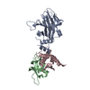 2fnjS S: Starting model for refinement C: citing same article ( |
|---|---|
| Similar structure data |


-Links
 PDBj
PDBj
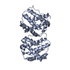
- Assembly
| Deposited unit | 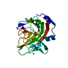
| ||||||||
|---|---|---|---|---|---|---|---|---|---|
| 1 | 
| ||||||||
| Unit cell |
| ||||||||
| Components on special symmetry positions |
|
-Components
| #1: Protein | Mass: 22756.725 Da / Num. of mol.: 1 / Fragment: RESIDUES 28-233 Source method: isolated from a genetically manipulated source Source: (gene. exp.) HOMO SAPIENS (human) / Plasmid: PNIC28-BSA4 / Production host:  |
|---|---|
| #2: Chemical | ChemComp-NI /   Mass: 58.693 Da / Num. of mol.: 1 / Source method: obtained synthetically / Formula: Ni Mass: 58.693 Da / Num. of mol.: 1 / Source method: obtained synthetically / Formula: Ni |
| #3: Water | ChemComp-HOH /  Mass: 18.015 Da / Num. of mol.: 78 / Source method: isolated from a natural source / Formula: H2O Mass: 18.015 Da / Num. of mol.: 78 / Source method: isolated from a natural source / Formula: H2O |
| Sequence details | THE FIRST TWO RESIDUES REMAIN FROM A CLEAVED HIS6 |
-Experimental details
-Experiment
| Experiment | Method: X-RAY DIFFRACTION / Number of used crystals: 1 |
|---|
- Sample preparation
| Crystal | Density Matthews: 2.7 Å3/Da / Density % sol: 55 % |
|---|---|
| Crystal grow | pH: 6.5 Details: 0.1 M NACACOD PH6.5, 14.4% PEG10K, 0.16 M CAAC2, 20% GLYCEROL, pH 6.50 |
-Data collection
| Diffraction | Mean temperature: 100 K |
|---|---|
| Diffraction source | Source: SYNCHROTRON / Site: SLS  / Beamline: X10SA / Wavelength: 1.03315 / Beamline: X10SA / Wavelength: 1.03315 |
| Detector | Type: MARRESEARCH / Detector: CCD / Date: Apr 15, 2007 |
| Radiation | Monochromator: SI111 / Protocol: SINGLE WAVELENGTH / Monochromatic (M) / Laue (L): M / Scattering type: x-ray |
| Radiation wavelength | Wavelength: 1.03315 Å / Relative weight: 1 |
| Reflection | Resolution: 2.2→45.1 Å / Num. obs: 13208 / % possible obs: 100 % / Observed criterion σ(I): 0 / Redundancy: 10.9 % / Rmerge(I) obs: 0.07 / Net I/σ(I): 18.7 |
| Reflection shell | Resolution: 2.2→2.3 Å / Redundancy: 10.9 % / Rmerge(I) obs: 0.56 / Mean I/σ(I) obs: 3.4 / % possible all: 99.9 |
- Processing
| Software |
| ||||||||||||||||||||||||||||||||||||||||||||||||||||||||||||||||||||||||||||||||||||||||||||||||||||||||||||||||||||||||||||||||||||||||||||||||||||||||||||||||||||||||||||||||||||||
|---|---|---|---|---|---|---|---|---|---|---|---|---|---|---|---|---|---|---|---|---|---|---|---|---|---|---|---|---|---|---|---|---|---|---|---|---|---|---|---|---|---|---|---|---|---|---|---|---|---|---|---|---|---|---|---|---|---|---|---|---|---|---|---|---|---|---|---|---|---|---|---|---|---|---|---|---|---|---|---|---|---|---|---|---|---|---|---|---|---|---|---|---|---|---|---|---|---|---|---|---|---|---|---|---|---|---|---|---|---|---|---|---|---|---|---|---|---|---|---|---|---|---|---|---|---|---|---|---|---|---|---|---|---|---|---|---|---|---|---|---|---|---|---|---|---|---|---|---|---|---|---|---|---|---|---|---|---|---|---|---|---|---|---|---|---|---|---|---|---|---|---|---|---|---|---|---|---|---|---|---|---|---|---|
| Refinement | Method to determine structure: MOLECULAR REPLACEMENT Starting model: PDB ENTRY 2FNJ Resolution: 2.2→50 Å / Cor.coef. Fo:Fc: 0.954 / Cor.coef. Fo:Fc free: 0.936 / SU B: 16.377 / SU ML: 0.212 / TLS residual ADP flag: UNVERIFIED / Cross valid method: THROUGHOUT / ESU R: 0.241 / ESU R Free: 0.209 / Stereochemistry target values: MAXIMUM LIKELIHOOD / Details: HYDROGENS HAVE BEEN ADDED IN THE RIDING POSITIONS.
| ||||||||||||||||||||||||||||||||||||||||||||||||||||||||||||||||||||||||||||||||||||||||||||||||||||||||||||||||||||||||||||||||||||||||||||||||||||||||||||||||||||||||||||||||||||||
| Solvent computation | Ion probe radii: 0.8 Å / Shrinkage radii: 0.8 Å / VDW probe radii: 1.2 Å / Solvent model: MASK | ||||||||||||||||||||||||||||||||||||||||||||||||||||||||||||||||||||||||||||||||||||||||||||||||||||||||||||||||||||||||||||||||||||||||||||||||||||||||||||||||||||||||||||||||||||||
| Displacement parameters | Biso mean: 23.05 Å2
| ||||||||||||||||||||||||||||||||||||||||||||||||||||||||||||||||||||||||||||||||||||||||||||||||||||||||||||||||||||||||||||||||||||||||||||||||||||||||||||||||||||||||||||||||||||||
| Refinement step | Cycle: LAST / Resolution: 2.2→50 Å
| ||||||||||||||||||||||||||||||||||||||||||||||||||||||||||||||||||||||||||||||||||||||||||||||||||||||||||||||||||||||||||||||||||||||||||||||||||||||||||||||||||||||||||||||||||||||
| Refine LS restraints |
|