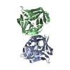
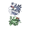
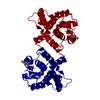
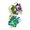
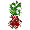
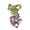
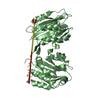
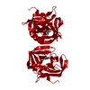
Entry Database : PDB / ID : 2vb0Title Crystal structure of coxsackievirus B3 proteinase 3C POLYPROTEIN 3BCD Keywords / / / / / / Function / homology Function Domain/homology Component
/ / / / / / / / / / / / / / / / / / / / / / / / / / / / / / / / / / / / / / / / / / / / / / / / / / / / / / / / / / / / / / / / / / / / / / / / / / / / / / / / / / / / / / / / / Biological species Method / / / Resolution : 2.4 Å Authors Anand, K. / Mesters, J.R. / Goerlach, R. / Zell, R. / Hilgenfeld, R. History Deposition Sep 5, 2007 Deposition site / Processing site Revision 1.0 Oct 28, 2008 Provider / Type Revision 1.1 Jul 13, 2011 Group / Version format complianceRevision 1.2 Jul 24, 2019 Group / Data collection / Derived calculationsCategory / pdbx_unobs_or_zero_occ_atoms / struct_connItem / _struct_conn.pdbx_leaving_atom_flagRevision 1.3 May 1, 2024 Group Advisory / Data collection ... Advisory / Data collection / Database references / Other / Refinement description Category chem_comp_atom / chem_comp_bond ... chem_comp_atom / chem_comp_bond / database_2 / pdbx_database_status / pdbx_initial_refinement_model / pdbx_unobs_or_zero_occ_atoms Item / _database_2.pdbx_database_accession / _pdbx_database_status.status_code_sfRevision 1.4 Nov 13, 2024 Group / Category / pdbx_modification_feature
Show all Show less Remark 700 SHEET DETERMINATION METHOD: DSSP THE SHEETS PRESENTED AS "AA" IN EACH CHAIN ON SHEET RECORDS BELOW ... SHEET DETERMINATION METHOD: DSSP THE SHEETS PRESENTED AS "AA" IN EACH CHAIN ON SHEET RECORDS BELOW IS ACTUALLY AN 7-STRANDED BARREL THIS IS REPRESENTED BY A 8-STRANDED SHEET IN WHICH THE FIRST AND LAST STRANDS ARE IDENTICAL. THE SHEETS PRESENTED AS "AB" IN EACH CHAIN ON SHEET RECORDS BELOW IS ACTUALLY AN 7-STRANDED BARREL THIS IS REPRESENTED BY A 8-STRANDED SHEET IN WHICH THE FIRST AND LAST STRANDS ARE IDENTICAL.
 Movie
Movie Controller


 Open data
 Basic information
 Components
 Keywords
 Function and homology information
 COXSACKIEVIRUS B3
 X-RAY DIFFRACTION /
  Authors
 Citation
 Structure visualization
 Downloads & links
 Other downloads
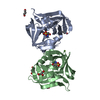


 PDBj
PDBj

 Assembly
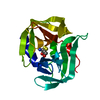


 Mass: 35.453 Da / Num. of mol.: 1 / Source method: obtained synthetically / Formula: Cl
 Mass: 18.015 Da / Num. of mol.: 47 / Source method: isolated from a natural source / Formula: H2O
 Sample preparation
 / Beamline: X13 / Wavelength: 0.804
  Processing