 Movie
Movie Controller


+ Open data

- Basic information
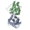
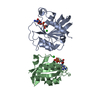
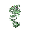
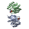
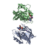
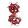
| Entry | Database: PDB / ID: 1gux | ||||||
|---|---|---|---|---|---|---|---|
| Title | RB POCKET BOUND TO E7 LXCXE MOTIF | ||||||
 Components Components |
| ||||||
 Keywords Keywords | COMPLEX (TRANSCRIPTION REG/PEPTIDE) / COMPLEX (TRANSCRIPTION REGULATION-PEPTIDE) / TUMOR SUPPRESSOR PROTEIN / RETINOBLASTOMA / COMPLEX (TRANSCRIPTION REG-PEPTIDE) COMPLEX | ||||||
| Function / homology |  Function and homology information Function and homology informationDefective translocation of RB1 mutants to the nucleus / enucleate erythrocyte differentiation / Rb-E2F complex / regulation of lipid kinase activity / positive regulation of collagen fibril organization / maintenance of mitotic sister chromatid cohesion / negative regulation of myofibroblast differentiation / chromatin lock complex / Positive Regulation of CDH1 Gene Transcription / cell morphogenesis involved in neuron differentiation ...Defective translocation of RB1 mutants to the nucleus / enucleate erythrocyte differentiation / Rb-E2F complex / regulation of lipid kinase activity / positive regulation of collagen fibril organization / maintenance of mitotic sister chromatid cohesion / negative regulation of myofibroblast differentiation / chromatin lock complex / Positive Regulation of CDH1 Gene Transcription / cell morphogenesis involved in neuron differentiation / sister chromatid biorientation / positive regulation of transcription regulatory region DNA binding / positive regulation of extracellular matrix organization / Aberrant regulation of mitotic exit in cancer due to RB1 defects / Inhibition of replication initiation of damaged DNA by RB1/E2F1 / glial cell apoptotic process / positive regulation of macrophage differentiation / negative regulation of hepatocyte apoptotic process / protein localization to chromosome, centromeric region / tissue homeostasis / importin-alpha family protein binding / neuron maturation / myoblast differentiation / positive regulation of mitotic metaphase/anaphase transition / Replication of the SARS-CoV-1 genome / aortic valve morphogenesis / digestive tract development / negative regulation of cold-induced thermogenesis / negative regulation of glial cell proliferation / SWI/SNF complex / Formation of Senescence-Associated Heterochromatin Foci (SAHF) / smoothened signaling pathway / negative regulation of G1/S transition of mitotic cell cycle / Phosphorylation of proteins involved in G1/S transition by active Cyclin E:Cdk2 complexes / hepatocyte apoptotic process / RUNX2 regulates osteoblast differentiation / Defective binding of RB1 mutants to E2F1,(E2F2, E2F3) / negative regulation of cell cycle / negative regulation of apoptotic signaling pathway / skeletal muscle cell differentiation / chondrocyte differentiation / chromosome organization / negative regulation of protein kinase activity / Cyclin E associated events during G1/S transition / Cyclin A:Cdk2-associated events at S phase entry / glial cell proliferation / striated muscle cell differentiation / Nuclear events stimulated by ALK signaling in cancer / epithelial cell proliferation / regulation of mitotic cell cycle / Condensation of Prophase Chromosomes / RNA polymerase II transcription regulatory region sequence-specific DNA binding / negative regulation of smoothened signaling pathway / phosphoprotein binding / G1/S transition of mitotic cell cycle / negative regulation of cell growth / PML body / APC/C:Cdh1 mediated degradation of Cdc20 and other APC/C:Cdh1 targeted proteins in late mitosis/early G1 / Oncogene Induced Senescence / negative regulation of inflammatory response / cellular response to xenobiotic stimulus / spindle / negative regulation of epithelial cell proliferation / kinase binding / cellular response to insulin stimulus / disordered domain specific binding / neuron projection development / Cyclin D associated events in G1 / transcription corepressor activity / heterochromatin formation / MLL4 and MLL3 complexes regulate expression of PPARG target genes in adipogenesis and hepatic steatosis / neuron apoptotic process / Replication of the SARS-CoV-2 genome / spermatogenesis / transcription by RNA polymerase II / DNA-binding transcription factor binding / molecular adaptor activity / RNA polymerase II-specific DNA-binding transcription factor binding / Ras protein signal transduction / cell differentiation / regulation of cell cycle / chromatin remodeling / negative regulation of gene expression / cell division / negative regulation of DNA-templated transcription / ubiquitin protein ligase binding / regulation of DNA-templated transcription / chromatin / negative regulation of transcription by RNA polymerase II / positive regulation of transcription by RNA polymerase II / nucleoplasm / identical protein binding / nucleus / cytoplasm / cytosol Similarity search - Function | ||||||
| Biological species |  Homo sapiens (human) Homo sapiens (human)  Human papillomavirus Human papillomavirus | ||||||
| Method |  X-RAY DIFFRACTION / SYNCHROTRON / MAD / Resolution: 1.85 Å X-RAY DIFFRACTION / SYNCHROTRON / MAD / Resolution: 1.85 Å | ||||||
 Authors Authors | Lee, J.O. / Russo, A.A. / Pavletich, N.P. | ||||||
 Citation Citation | Journal: Nature / Year: 1998 Title: Structure of the retinoblastoma tumour-suppressor pocket domain bound to a peptide from HPV E7. Authors: Lee, J.O. / Russo, A.A. / Pavletich, N.P. | ||||||
| History |
|
- Structure visualization
| Structure viewer | Molecule: MolmilJmol/JSmol |
|---|
- Downloads & links
-Download
| PDBx/mmCIF format | 1gux.cif.gz | 88.7 KB | Display | PDBx/mmCIF format |
|---|---|---|---|---|
| PDB format | pdb1gux.ent.gz | 67 KB | Display | PDB format |
| PDBx/mmJSON format | 1gux.json.gz | Tree view | PDBx/mmJSON format | |
| Others |  Other downloads Other downloads |
-Validation report
| Arichive directory | https://data.pdbj.org/pub/pdb/validation_reports/gu/1guxftp://data.pdbj.org/pub/pdb/validation_reports/gu/1gux | HTTPS FTP |
|---|
-Related structure data
| Similar structure data |
|---|


-Links
 PDBj
PDBj


- Assembly
| Deposited unit | 
| ||||||||
|---|---|---|---|---|---|---|---|---|---|
| 1 |
| ||||||||
| Unit cell |
|
-Components
| #1: Protein | Mass: 25100.910 Da / Num. of mol.: 1 / Fragment: POCKET DOMAIN Source method: isolated from a genetically manipulated source Source: (gene. exp.) Homo sapiens (human) / Cell line: BL21 / Plasmid: BL21 / Species (production host): Escherichia coli / Production host:  |
|---|---|
| #2: Protein | Mass: 18265.477 Da / Num. of mol.: 1 / Fragment: POCKET DOMAIN Source method: isolated from a genetically manipulated source Source: (gene. exp.) Homo sapiens (human) / Cell line: BL21 / Plasmid: BL21 / Species (production host): Escherichia coli / Production host: |
| #3: Protein/peptide | Mass: 1160.255 Da / Num. of mol.: 1 / Source method: isolated from a natural source Details: THE NINE RESIDUES OF PAPILLOMA VIRUS E7 PEPTIDE CONTAIN THE LXCXE MOTIF Source: (natural) Human papillomavirus / Strain: E7 |
| #4: Water | ChemComp-HOH /  Mass: 18.015 Da / Num. of mol.: 388 / Source method: isolated from a natural source / Formula: H2O Mass: 18.015 Da / Num. of mol.: 388 / Source method: isolated from a natural source / Formula: H2O |
-Experimental details
-Experiment
| Experiment | Method: X-RAY DIFFRACTION / Number of used crystals: 1 |
|---|
- Sample preparation
| Crystal | Density Matthews: 2.86 Å3/Da / Density % sol: 56.92 % | ||||||||||||||||||||||||
|---|---|---|---|---|---|---|---|---|---|---|---|---|---|---|---|---|---|---|---|---|---|---|---|---|---|
| Crystal grow | pH: 7.5 Details: 3.5 M SODIUM FORMATE, 0.1M NA HEPES PH 7.5, 20 MIROMOLAR CDCL2 | ||||||||||||||||||||||||
| Crystal grow | *PLUS Temperature: 4 ℃ / Method: vapor diffusion, hanging drop | ||||||||||||||||||||||||
| Components of the solutions | *PLUS
|
-Data collection
| Diffraction | Mean temperature: 100 K |
|---|---|
| Diffraction source | Source: SYNCHROTRON / Site: CHESS  / Beamline: A1 / Wavelength: 0.908 / Beamline: A1 / Wavelength: 0.908 |
| Detector | Type: ADSC QUANTUM / Detector: CCD / Date: Jul 7, 1997 |
| Radiation | Monochromatic (M) / Laue (L): M / Scattering type: x-ray |
| Radiation wavelength | Wavelength: 0.908 Å / Relative weight: 1 |
| Reflection | Highest resolution: 1.85 Å / Num. obs: 45533 / % possible obs: 91.8 % / Observed criterion σ(I): 0 / Redundancy: 2.98 % / Rsym value: 0.048 / Net I/σ(I): 25.1 |
| Reflection shell | Resolution: 1.85→1.94 Å / Redundancy: 1.98 % / Mean I/σ(I) obs: 5.93 / Rsym value: 0.211 / % possible all: 84.3 |
| Reflection | *PLUS Num. measured all: 135820 / Rmerge(I) obs: 0.048 |
| Reflection shell | *PLUS % possible obs: 84.3 % / Rmerge(I) obs: 0.211 |
- Processing
| Software |
| ||||||||||||||||||||||||||||||||||||||||||||||||||||||||||||
|---|---|---|---|---|---|---|---|---|---|---|---|---|---|---|---|---|---|---|---|---|---|---|---|---|---|---|---|---|---|---|---|---|---|---|---|---|---|---|---|---|---|---|---|---|---|---|---|---|---|---|---|---|---|---|---|---|---|---|---|---|---|
| Refinement | Method to determine structure: MAD / Resolution: 1.85→7 Å / σ(F): 2
| ||||||||||||||||||||||||||||||||||||||||||||||||||||||||||||
| Displacement parameters | Biso mean: 31.6 Å2 | ||||||||||||||||||||||||||||||||||||||||||||||||||||||||||||
| Refinement step | Cycle: LAST / Resolution: 1.85→7 Å
| ||||||||||||||||||||||||||||||||||||||||||||||||||||||||||||
| Refine LS restraints |
| ||||||||||||||||||||||||||||||||||||||||||||||||||||||||||||
| LS refinement shell | Resolution: 1.85→1.93 Å / Total num. of bins used: 8
| ||||||||||||||||||||||||||||||||||||||||||||||||||||||||||||
| Software | *PLUS Name: X-PLOR / Version: 3.1 / Classification: refinement | ||||||||||||||||||||||||||||||||||||||||||||||||||||||||||||
| Refinement | *PLUS | ||||||||||||||||||||||||||||||||||||||||||||||||||||||||||||
| Solvent computation | *PLUS | ||||||||||||||||||||||||||||||||||||||||||||||||||||||||||||
| Displacement parameters | *PLUS | ||||||||||||||||||||||||||||||||||||||||||||||||||||||||||||
| Refine LS restraints | *PLUS
| ||||||||||||||||||||||||||||||||||||||||||||||||||||||||||||
| LS refinement shell | *PLUS Rfactor obs: 0.324 |
