 Movie
Movie Controller

[English] 日本語
 Yorodumi
- PDB-1n4m: Structure of Rb tumor suppressor bound to the transactivation dom... -
+ Open data

- Basic information
| Entry | Database: PDB / ID: 1n4m | ||||||
|---|---|---|---|---|---|---|---|
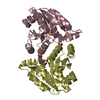
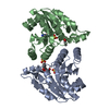
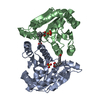
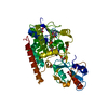
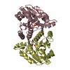
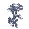
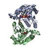
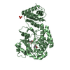
| Title | Structure of Rb tumor suppressor bound to the transactivation domain of E2F-2 | ||||||
 Components Components |
| ||||||
 Keywords Keywords | CELL CYCLE / PROTEIN-PEPTIDE COMPLEX | ||||||
| Function / homology |  Function and homology information Function and homology informationDefective translocation of RB1 mutants to the nucleus / Rb-E2F complex / regulation of lipid kinase activity / positive regulation of collagen fibril organization / maintenance of mitotic sister chromatid cohesion / negative regulation of myofibroblast differentiation / negative regulation of sprouting angiogenesis / cell morphogenesis involved in neuron differentiation / Positive Regulation of CDH1 Gene Transcription / sister chromatid biorientation ...Defective translocation of RB1 mutants to the nucleus / Rb-E2F complex / regulation of lipid kinase activity / positive regulation of collagen fibril organization / maintenance of mitotic sister chromatid cohesion / negative regulation of myofibroblast differentiation / negative regulation of sprouting angiogenesis / cell morphogenesis involved in neuron differentiation / Positive Regulation of CDH1 Gene Transcription / sister chromatid biorientation / positive regulation of transcription regulatory region DNA binding / positive regulation of extracellular matrix organization / Aberrant regulation of mitotic exit in cancer due to RB1 defects / negative regulation of hepatocyte apoptotic process / Inhibition of replication initiation of damaged DNA by RB1/E2F1 / protein localization to chromosome, centromeric region / importin-alpha family protein binding / myoblast differentiation / positive regulation of mitotic metaphase/anaphase transition / Replication of the SARS-CoV-1 genome / aortic valve morphogenesis / negative regulation of cold-induced thermogenesis / SWI/SNF complex / Formation of Senescence-Associated Heterochromatin Foci (SAHF) / negative regulation of G1/S transition of mitotic cell cycle / Phosphorylation of proteins involved in G1/S transition by active Cyclin E:Cdk2 complexes / RUNX2 regulates osteoblast differentiation / Defective binding of RB1 mutants to E2F1,(E2F2, E2F3) / negative regulation of cell cycle / chondrocyte differentiation / negative regulation of apoptotic signaling pathway / chromosome organization / Cyclin E associated events during G1/S transition / negative regulation of protein kinase activity / Cyclin A:Cdk2-associated events at S phase entry / cis-regulatory region sequence-specific DNA binding / Nuclear events stimulated by ALK signaling in cancer / regulation of mitotic cell cycle / Condensation of Prophase Chromosomes / RNA polymerase II transcription regulatory region sequence-specific DNA binding / transcription initiation at RNA polymerase II promoter / phosphoprotein binding / negative regulation of cell growth / negative regulation of inflammatory response / PML body / Oncogene Induced Senescence / APC/C:Cdh1 mediated degradation of Cdc20 and other APC/C:Cdh1 targeted proteins in late mitosis/early G1 / RNA polymerase II transcription regulator complex / spindle / kinase binding / neuron projection development / cellular response to insulin stimulus / disordered domain specific binding / sequence-specific double-stranded DNA binding / Cyclin D associated events in G1 / transcription corepressor activity / heterochromatin formation / MLL4 and MLL3 complexes regulate expression of PPARG target genes in adipogenesis and hepatic steatosis / DNA-binding transcription activator activity, RNA polymerase II-specific / Replication of the SARS-CoV-2 genome / Oxidative Stress Induced Senescence / DNA-binding transcription factor binding / spermatogenesis / sequence-specific DNA binding / molecular adaptor activity / RNA polymerase II-specific DNA-binding transcription factor binding / DNA-binding transcription factor activity, RNA polymerase II-specific / Ras protein signal transduction / cell differentiation / regulation of cell cycle / protein dimerization activity / RNA polymerase II cis-regulatory region sequence-specific DNA binding / chromatin remodeling / DNA-binding transcription factor activity / negative regulation of gene expression / negative regulation of DNA-templated transcription / apoptotic process / ubiquitin protein ligase binding / regulation of transcription by RNA polymerase II / regulation of DNA-templated transcription / chromatin / negative regulation of transcription by RNA polymerase II / DNA-templated transcription / positive regulation of transcription by RNA polymerase II / DNA binding / nucleoplasm / identical protein binding / nucleus / cytosol / cytoplasm Similarity search - Function | ||||||
| Biological species |  Homo sapiens (human) Homo sapiens (human) | ||||||
| Method |  X-RAY DIFFRACTION / SYNCHROTRON / MOLECULAR REPLACEMENT / Resolution: 2.2 Å X-RAY DIFFRACTION / SYNCHROTRON / MOLECULAR REPLACEMENT / Resolution: 2.2 Å | ||||||
 Authors Authors | Lee, C. / Chang, J.H. / Lee, H.S. / Cho, Y. | ||||||
 Citation Citation | Journal: GENES DEV. / Year: 2002 Title: Structural basis for the recognition of the E2F transactivation domain by the retinoblastoma tumor suppressor Authors: Lee, C. / Chang, J.H. / Lee, H.S. / Cho, Y. | ||||||
| History |
|
- Structure visualization
| Structure viewer | Molecule: MolmilJmol/JSmol |
|---|
- Downloads & links
-Download
| PDBx/mmCIF format | 1n4m.cif.gz | 161 KB | Display | PDBx/mmCIF format |
|---|---|---|---|---|
| PDB format | pdb1n4m.ent.gz | 127.9 KB | Display | PDB format |
| PDBx/mmJSON format | 1n4m.json.gz | Tree view | PDBx/mmJSON format | |
| Others |  Other downloads Other downloads |
-Validation report
| Arichive directory | https://data.pdbj.org/pub/pdb/validation_reports/n4/1n4mftp://data.pdbj.org/pub/pdb/validation_reports/n4/1n4m | HTTPS FTP |
|---|
-Related structure data
| Similar structure data |
|---|

-Links
 PDBj
PDBj


- Assembly
| Deposited unit | 
| ||||||||
|---|---|---|---|---|---|---|---|---|---|
| 1 | 
| ||||||||
| 2 | 
| ||||||||
| 3 | 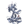
| ||||||||
| 4 | 
| ||||||||
| Unit cell |
|
-Components
| #1: Protein | Mass: 40344.926 Da / Num. of mol.: 2 / Fragment: Residues 380-785 Source method: isolated from a genetically manipulated source Source: (gene. exp.) Homo sapiens (human) / Plasmid: pGEX4T3 / Production host:  #2: Protein/peptide | Mass: 2015.094 Da / Num. of mol.: 3 / Fragment: Residues 410-427 / Source method: obtained synthetically Details: The peptide was chemically synthesized. The sequence of the peptide is naturally found in Homo Sapiens (human). References: UniProt: Q14209 #3: Water | ChemComp-HOH / |  Mass: 18.015 Da / Num. of mol.: 261 / Source method: isolated from a natural source / Formula: H2O Mass: 18.015 Da / Num. of mol.: 261 / Source method: isolated from a natural source / Formula: H2O |
|---|
-Experimental details
-Experiment
| Experiment | Method: X-RAY DIFFRACTION / Number of used crystals: 1 |
|---|
- Sample preparation
| Crystal | Density Matthews: 2.57 Å3/Da / Density % sol: 52.06 % | |||||||||||||||||||||||||||||||||||||||||||||||||
|---|---|---|---|---|---|---|---|---|---|---|---|---|---|---|---|---|---|---|---|---|---|---|---|---|---|---|---|---|---|---|---|---|---|---|---|---|---|---|---|---|---|---|---|---|---|---|---|---|---|---|
| Crystal grow | Temperature: 277 K / Method: vapor diffusion, hanging drop / pH: 5.5 Details: Ammonium sulfate Citrate, pH 5.5, VAPOR DIFFUSION, HANGING DROP, temperature 277K | |||||||||||||||||||||||||||||||||||||||||||||||||
| Crystal grow | *PLUS Temperature: 4 ℃ / pH: 7.5 | |||||||||||||||||||||||||||||||||||||||||||||||||
| Components of the solutions | *PLUS
|
-Data collection
| Diffraction source | Source: SYNCHROTRON / Site: PAL/PLS  / Beamline: 6B / Wavelength: 1.12714 Å / Beamline: 6B / Wavelength: 1.12714 Å |
|---|---|
| Detector | Type: MACSCIENCE / Detector: IMAGE PLATE / Date: Jun 2, 2002 |
| Radiation | Protocol: SINGLE WAVELENGTH / Monochromatic (M) / Laue (L): M / Scattering type: x-ray |
| Radiation wavelength | Wavelength: 1.12714 Å / Relative weight: 1 |
| Reflection | Resolution: 2.2→50 Å / Num. obs: 37599 / Observed criterion σ(I): 1 / Biso Wilson estimate: 4.8 Å2 |
| Reflection | *PLUS Lowest resolution: 50 Å / % possible obs: 86.3 % / Num. measured all: 93798 / Rmerge(I) obs: 0.082 |
| Reflection shell | *PLUS Highest resolution: 2.2 Å / Lowest resolution: 2.28 Å / % possible obs: 70.3 % / Rmerge(I) obs: 0.237 |
- Processing
| Software |
| ||||||||||||||||||||||||||||||||||||||||||||||||||||||||||||||||||||||||||||||||
|---|---|---|---|---|---|---|---|---|---|---|---|---|---|---|---|---|---|---|---|---|---|---|---|---|---|---|---|---|---|---|---|---|---|---|---|---|---|---|---|---|---|---|---|---|---|---|---|---|---|---|---|---|---|---|---|---|---|---|---|---|---|---|---|---|---|---|---|---|---|---|---|---|---|---|---|---|---|---|---|---|---|
| Refinement | Method to determine structure: MOLECULAR REPLACEMENT / Resolution: 2.2→19.65 Å / Rfactor Rfree error: 0.007 / Data cutoff high absF: 369771.74 / Data cutoff high rms absF: 369771.74 / Data cutoff low absF: 0 / Isotropic thermal model: RESTRAINED / Cross valid method: THROUGHOUT / σ(F): 0
| ||||||||||||||||||||||||||||||||||||||||||||||||||||||||||||||||||||||||||||||||
| Solvent computation | Solvent model: FLAT MODEL / Bsol: 45.9332 Å2 / ksol: 0.390844 e/Å3 | ||||||||||||||||||||||||||||||||||||||||||||||||||||||||||||||||||||||||||||||||
| Displacement parameters | Biso mean: 25.4 Å2
| ||||||||||||||||||||||||||||||||||||||||||||||||||||||||||||||||||||||||||||||||
| Refine analyze |
| ||||||||||||||||||||||||||||||||||||||||||||||||||||||||||||||||||||||||||||||||
| Refinement step | Cycle: LAST / Resolution: 2.2→19.65 Å
| ||||||||||||||||||||||||||||||||||||||||||||||||||||||||||||||||||||||||||||||||
| Refine LS restraints |
| ||||||||||||||||||||||||||||||||||||||||||||||||||||||||||||||||||||||||||||||||
| LS refinement shell | Resolution: 2.2→2.34 Å / Rfactor Rfree error: 0.021 / Total num. of bins used: 6
| ||||||||||||||||||||||||||||||||||||||||||||||||||||||||||||||||||||||||||||||||
| Xplor file |
| ||||||||||||||||||||||||||||||||||||||||||||||||||||||||||||||||||||||||||||||||
| Refinement | *PLUS Highest resolution: 2.2 Å / Lowest resolution: 20 Å / % reflection Rfree: 5 % / Rfactor Rfree: 0.286 / Rfactor Rwork: 0.224 | ||||||||||||||||||||||||||||||||||||||||||||||||||||||||||||||||||||||||||||||||
| Solvent computation | *PLUS | ||||||||||||||||||||||||||||||||||||||||||||||||||||||||||||||||||||||||||||||||
| Displacement parameters | *PLUS | ||||||||||||||||||||||||||||||||||||||||||||||||||||||||||||||||||||||||||||||||
| Refine LS restraints | *PLUS
| ||||||||||||||||||||||||||||||||||||||||||||||||||||||||||||||||||||||||||||||||
| LS refinement shell | *PLUS Lowest resolution: 2.28 Å / Rfactor Rfree: 0.328 / Rfactor Rwork: 0.287 |
