 Movie
Movie Controller

[English] 日本語
 Yorodumi
- PDB-3oc5: Crystal Structure of the vibrio cholerae secreted colonization fa... -
+ Open data

- Basic information
| Entry | Database: PDB / ID: 3oc5 | ||||||
|---|---|---|---|---|---|---|---|
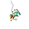
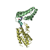
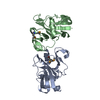
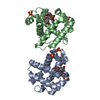
| Title | Crystal Structure of the vibrio cholerae secreted colonization factor TcpF | ||||||
 Components Components | Toxin coregulated pilus biosynthesis protein F | ||||||
 Keywords Keywords | CELL ADHESION / MULTIDOMAIN PROTEIN / immunoglobulin-like (Ig-like) fold | ||||||
| Function / homology | Vibrio cholerae toxin co-regulated pilus biosynthesis protein F, C-terminal domain / Vibrio cholerae toxin co-regulated pilus biosynthesis F / Vibrio cholerae toxin co-regulated pilus biosynthesis F, C-terminal / Vibrio cholerae toxin co-regulated pilus biosynthesis protein F / cell outer membrane / Immunoglobulin-like / Sandwich / Mainly Beta / Toxin coregulated pilus biosynthesis protein F Function and homology information Function and homology information | ||||||
| Biological species |   Vibrio cholerae (bacteria) Vibrio cholerae (bacteria) | ||||||
| Method |  X-RAY DIFFRACTION / SYNCHROTRON / MAD / Resolution: 2.4 Å X-RAY DIFFRACTION / SYNCHROTRON / MAD / Resolution: 2.4 Å | ||||||
 Authors Authors | Craig, L. / Kolappan, S. / Yuen, A.S.W. | ||||||
 Citation Citation | Journal: J.Mol.Biol. / Year: 2011 Title: Crystal Structure of the Vibrio cholerae Colonization Factor TcpF and Identification of a Functional Immunogenic Site. Authors: Megli, C.J. / Yuen, A.S. / Kolappan, S. / Richardson, M.R. / Dharmasena, M.N. / Krebs, S.J. / Taylor, R.K. / Craig, L. | ||||||
| History |
|
- Structure visualization
| Structure viewer | Molecule: MolmilJmol/JSmol |
|---|
- Downloads & links
-Download
| PDBx/mmCIF format | 3oc5.cif.gz | 125.8 KB | Display | PDBx/mmCIF format |
|---|---|---|---|---|
| PDB format | pdb3oc5.ent.gz | 100.7 KB | Display | PDB format |
| PDBx/mmJSON format | 3oc5.json.gz | Tree view | PDBx/mmJSON format | |
| Others |  Other downloads Other downloads |
-Validation report
| Arichive directory | https://data.pdbj.org/pub/pdb/validation_reports/oc/3oc5ftp://data.pdbj.org/pub/pdb/validation_reports/oc/3oc5 | HTTPS FTP |
|---|
-Related structure data


-Links
 PDBj
PDBj- Assembly
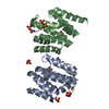
| Deposited unit | 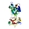
| ||||||||
|---|---|---|---|---|---|---|---|---|---|
| 1 |
| ||||||||
| 2 | x 12
| ||||||||
| Unit cell |
|
-Components
| #1: Protein | Mass: 35882.938 Da / Num. of mol.: 1 / Fragment: UNP residues 21-338 Source method: isolated from a genetically manipulated source Source: (gene. exp.) Vibrio cholerae (bacteria) / Gene: tcpF, VC_0837 / Production host: Vibrio cholerae (bacteria) / References: UniProt: P0C6Q5 |
|---|---|
| #2: Water | ChemComp-HOH /  Mass: 18.015 Da / Num. of mol.: 132 / Source method: isolated from a natural source / Formula: H2O Mass: 18.015 Da / Num. of mol.: 132 / Source method: isolated from a natural source / Formula: H2O |
| Has protein modification | Y |
-Experimental details
-Experiment
| Experiment | Method: X-RAY DIFFRACTION / Number of used crystals: 1 |
|---|
- Sample preparation
| Crystal | Density Matthews: 3.3 Å3/Da / Density % sol: 62.74 % |
|---|---|
| Crystal grow | Temperature: 293 K / pH: 7.5 Details: (NH4)2 H PO4 1M, Imidazole 100 mM, pH 7.5, VAPOR DIFFUSION, HANGING DROP, temperature 293K |
-Data collection
| Diffraction | Mean temperature: 100 K | |||||||||
|---|---|---|---|---|---|---|---|---|---|---|
| Diffraction source | Source: SYNCHROTRON / Site: ALS  / Beamline: 8.2.1 / Wavelength: 0.9116,0.9794 / Beamline: 8.2.1 / Wavelength: 0.9116,0.9794 | |||||||||
| Detector | Type: ADSC QUANTUM 315r / Detector: CCD / Date: Jul 15, 2007 | |||||||||
| Radiation | Monochromator: SI 111 / Protocol: MAD / Monochromatic (M) / Laue (L): M / Scattering type: x-ray | |||||||||
| Radiation wavelength |
| |||||||||
| Reflection | Resolution: 2.4→20 Å / Num. obs: 19524 / % possible obs: 99.4 % / Observed criterion σ(I): 2.3 |
- Processing
| Software |
| ||||||||||||||||||||||||||||||||||||||||||||||||||||||||||||||||||||||||||||||||||||||||||||||||||||||||||||||||||||||||||||||||||||||||||||||||||||||||||||||||||||||||||
|---|---|---|---|---|---|---|---|---|---|---|---|---|---|---|---|---|---|---|---|---|---|---|---|---|---|---|---|---|---|---|---|---|---|---|---|---|---|---|---|---|---|---|---|---|---|---|---|---|---|---|---|---|---|---|---|---|---|---|---|---|---|---|---|---|---|---|---|---|---|---|---|---|---|---|---|---|---|---|---|---|---|---|---|---|---|---|---|---|---|---|---|---|---|---|---|---|---|---|---|---|---|---|---|---|---|---|---|---|---|---|---|---|---|---|---|---|---|---|---|---|---|---|---|---|---|---|---|---|---|---|---|---|---|---|---|---|---|---|---|---|---|---|---|---|---|---|---|---|---|---|---|---|---|---|---|---|---|---|---|---|---|---|---|---|---|---|---|---|---|---|---|
| Refinement | Method to determine structure: MAD / Resolution: 2.4→19.65 Å / Cor.coef. Fo:Fc: 0.928 / Cor.coef. Fo:Fc free: 0.904 / SU B: 14.281 / SU ML: 0.153 / Cross valid method: THROUGHOUT / σ(F): 2.3 / ESU R Free: 0.22 / Stereochemistry target values: MAXIMUM LIKELIHOOD / Details: HYDROGENS HAVE BEEN ADDED IN THE RIDING POSITIONS
| ||||||||||||||||||||||||||||||||||||||||||||||||||||||||||||||||||||||||||||||||||||||||||||||||||||||||||||||||||||||||||||||||||||||||||||||||||||||||||||||||||||||||||
| Solvent computation | Ion probe radii: 0.8 Å / Shrinkage radii: 0.8 Å / VDW probe radii: 1.4 Å / Solvent model: MASK | ||||||||||||||||||||||||||||||||||||||||||||||||||||||||||||||||||||||||||||||||||||||||||||||||||||||||||||||||||||||||||||||||||||||||||||||||||||||||||||||||||||||||||
| Displacement parameters | Biso mean: 58.62 Å2 | ||||||||||||||||||||||||||||||||||||||||||||||||||||||||||||||||||||||||||||||||||||||||||||||||||||||||||||||||||||||||||||||||||||||||||||||||||||||||||||||||||||||||||
| Refinement step | Cycle: LAST / Resolution: 2.4→19.65 Å
| ||||||||||||||||||||||||||||||||||||||||||||||||||||||||||||||||||||||||||||||||||||||||||||||||||||||||||||||||||||||||||||||||||||||||||||||||||||||||||||||||||||||||||
| Refine LS restraints |
| ||||||||||||||||||||||||||||||||||||||||||||||||||||||||||||||||||||||||||||||||||||||||||||||||||||||||||||||||||||||||||||||||||||||||||||||||||||||||||||||||||||||||||
| LS refinement shell | Resolution: 2.4→2.46 Å / Total num. of bins used: 20
| ||||||||||||||||||||||||||||||||||||||||||||||||||||||||||||||||||||||||||||||||||||||||||||||||||||||||||||||||||||||||||||||||||||||||||||||||||||||||||||||||||||||||||
| Refinement TLS params. | Method: refined / Origin x: 59.423 Å / Origin y: -38.2792 Å / Origin z: -15.7493 Å
|
