 ムービー
ムービー コントローラー
 構造ビューア
 EMN検索について


-検索条件
-検索結果
検索 (著者・登録者: guerra & m)の結果全42件を表示しています

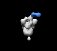
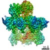
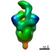
EMDB-18182: 
Closed conformation of the g-tubulin ring complex nucleating microtubules
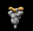
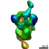
EMDB-18181:
Early closed conformation of the g-tubulin ring complex
PDB-8q62:
Early closed conformation of the g-tubulin ring complex
EMDB-28850:
SARS-CoV-2 Gamma 6P Mut7 S + COVA309-3 Fab
EMDB-28851:
SARS-CoV-2 Gamma 6P Mut7 S + COVA309-10 Fab
EMDB-28852:
SARS-CoV-2 Omicron 6P S + COVA309-35 Fab
EMDB-28853:
SARS-CoV-2 Gamma 6P Mut7 + S COVA309-38 Fab

EMDB-27177:
sd1.040 Fab in complex with SARS-CoV-2 Spike 2P glycoprotein

PDB-8d48:
sd1.040 Fab in complex with SARS-CoV-2 Spike 2P glycoprotein
EMDB-25412:
Structure of the human COQ7:COQ9 complex by single-particle electron cryo-microscopy, unliganded state
EMDB-25413:
Structure of the NADH-bound human COQ7:COQ9 complex by single-particle electron cryo-microscopy

PDB-7ssp:
Structure of the human COQ7:COQ9 complex by single-particle electron cryo-microscopy, unliganded state

PDB-7sss:
Structure of the NADH-bound human COQ7:COQ9 complex by single-particle electron cryo-microscopy
EMDB-26217:
Negative stain EM map of COVA1-07 mAb bound to the S2 domain of SARS-CoV-2 S
EMDB-26218:
Negative stain EM map of COVA2-14 mAb bound to the S2 domain of SARS-CoV-2 S
EMDB-26219:
Negative stain EM map of COVA2-18 mAb bound to the S2 domain of SARS-CoV-2 S
EMDB-26220:
Negative stain EM map of the S2 domain of SARS-CoV-2 S

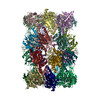
EMDB-13482:
Half-vault structure

PDB-7pky:
Half-vault structure

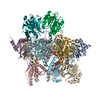
EMDB-13478:
Vault structure in primmed conformation

EMDB-13483:
Vault structure in committed conformation
PDB-7pkr:
Vault structure in primmed conformation

PDB-7pkz:
Vault structure in committed conformation

PDB-7ko8:
Cryo-EM structure of the mature and infective Mayaro virus
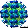
EMDB-22961:
Cryo-EM structure of the mature and infective Mayaro virus
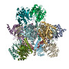
EMDB-23502:
Cryo-EM structure of the Pre3-1 20S proteasome core particle
EMDB-23503:
Cryo-EM structure of Pre-15S proteasome core particle assembly intermediate purified from Pre3-1 proteasome mutant (G34D)
EMDB-23508:
Cryo-EM structure of 13S proteasome core particle assembly intermediate purified from Pre3-1 proteasome mutant (G34D)
PDB-7ls5:
Cryo-EM structure of the Pre3-1 20S proteasome core particle
PDB-7ls6:
Cryo-EM structure of Pre-15S proteasome core particle assembly intermediate purified from Pre3-1 proteasome mutant (G34D)
PDB-7lsx:
Cryo-EM structure of 13S proteasome core particle assembly intermediate purified from Pre3-1 proteasome mutant (G34D)

EMDB-11236:
Reconstruction of Tula virus surface glycoprotein lattice

PDB-6zjm:
Atomic model of Andes virus glycoprotein spike tetramer generated by fitting into a Tula virus reconstruction
EMDB-22061:
negative stain EM map of SARS-CoV-2 spike in complex with COVA2-15 Fab
EMDB-22062:
negative stain EM map of SARS-CoV-2 spike in complex with COVA1-22
EMDB-22063:
negative stain EM map of SARS-CoV-2 spike in complex with COVA2-07
EMDB-22064:
negative stain EM map of SARS-CoV-2 spike in complex with COVA2-39 Fab
EMDB-22065:
negative stain EM map of SARS-CoV-2 spike in complex with COVA2-04 Fab
EMDB-22066:
negative stain EM map of SARS-CoV-2 spike in complex with COVA1-12 Fab

EMDB-0340:
EM structure of the DNA wrapping in bacterial open transcription initiation complex
EMDB-4867:
Sub-tomogram averaging of Tula virus glycoprotein spike

EMDB-2101:
Electron tomography of forming Hepatitis C virus-like particles in insect cells.
 wwPDBはEMDBデータモデルのバージョン3へ移行します
