National Institutes of Health/National Institute of General Medical Sciences (NIH/NIGMS)
5R01GM127673-04
United States
Citation
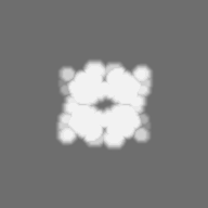
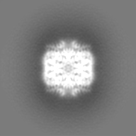
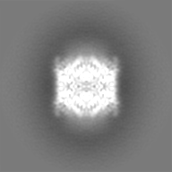
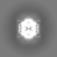
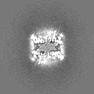
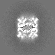
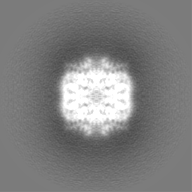
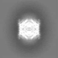
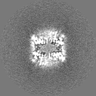
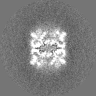
Journal: Mol Cell / Year: 2022 Title: Structure and functionality of a multimeric human COQ7:COQ9 complex. Authors: Mateusz Manicki / Halil Aydin / Luciano A Abriata / Katherine A Overmyer / Rachel M Guerra / Joshua J Coon / Matteo Dal Peraro / Adam Frost / David J Pagliarini / Abstract: Coenzyme Q (CoQ) is a redox-active lipid essential for core metabolic pathways and antioxidant defense. CoQ is synthesized upon the mitochondrial inner membrane by an ill-defined "complex Q" ...Coenzyme Q (CoQ) is a redox-active lipid essential for core metabolic pathways and antioxidant defense. CoQ is synthesized upon the mitochondrial inner membrane by an ill-defined "complex Q" metabolon. Here, we present structure-function analyses of a lipid-, substrate-, and NADH-bound complex comprising two complex Q subunits: the hydroxylase COQ7 and the lipid-binding protein COQ9. We reveal that COQ7 adopts a ferritin-like fold with a hydrophobic channel whose substrate-binding capacity is enhanced by COQ9. Using molecular dynamics, we further show that two COQ7:COQ9 heterodimers form a curved tetramer that deforms the membrane, potentially opening a pathway for the CoQ intermediates to translocate from the bilayer to the proteins' lipid-binding sites. Two such tetramers assemble into a soluble octamer with a pseudo-bilayer of lipids captured within. Together, these observations indicate that COQ7 and COQ9 cooperate to access hydrophobic precursors within the membrane and coordinate subsequent synthesis steps toward producing CoQ.
In the structure databanks used in Yorodumi, some data are registered as the other names, "COVID-19 virus" and "2019-nCoV". Here are the details of the virus and the list of structure data.
Jan 31, 2019. EMDB accession codes are about to change! (news from PDBe EMDB page)
EMDB accession codes are about to change! (news from PDBe EMDB page)
The allocation of 4 digits for EMDB accession codes will soon come to an end. Whilst these codes will remain in use, new EMDB accession codes will include an additional digit and will expand incrementally as the available range of codes is exhausted. The current 4-digit format prefixed with “EMD-” (i.e. EMD-XXXX) will advance to a 5-digit format (i.e. EMD-XXXXX), and so on. It is currently estimated that the 4-digit codes will be depleted around Spring 2019, at which point the 5-digit format will come into force.
The EM Navigator/Yorodumi systems omit the EMD- prefix.
Related info.:Q: What is EMD? / ID/Accession-code notation in Yorodumi/EM Navigator
Yorodumi is a browser for structure data from EMDB, PDB, SASBDB, etc.
This page is also the successor to EM Navigator detail page, and also detail information page/front-end page for Omokage search.
The word "yorodu" (or yorozu) is an old Japanese word meaning "ten thousand". "mi" (miru) is to see.
Related info.:EMDB / PDB / SASBDB / Comparison of 3 databanks / Yorodumi Search / Aug 31, 2016. New EM Navigator & Yorodumi / Yorodumi Papers / Jmol/JSmol / Function and homology information / Changes in new EM Navigator and Yorodumi
 Movie
Movie Controller

 Yorodumi
 Open data
 Basic information
 Map data
 Sample
 Keywords
 Function and homology information
 Homo sapiens (human)
 Authors
 United States, 1 items
  Citation
 Structure visualization
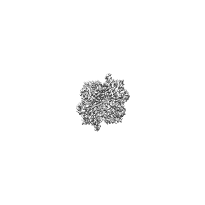
 Downloads & links
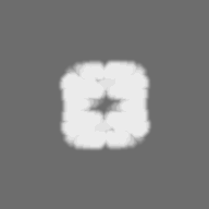
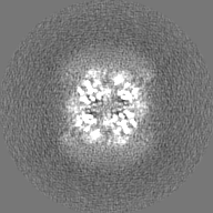
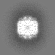
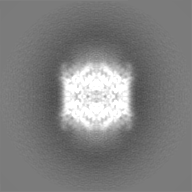
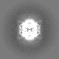
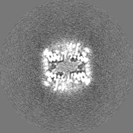
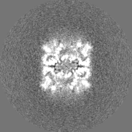
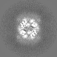
 emd_25412.png
 http://ftp.pdbj.org/pub/emdb/structures/EMD-25412


 Z (Sec.)
 Y (Row.)
 X (Col.)


 Sample components
 Processing
 Electron microscopy
 FIELD EMISSION GUN