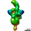
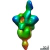
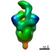
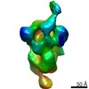
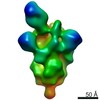
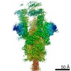
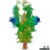
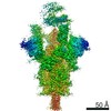
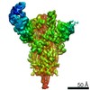
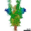
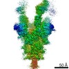
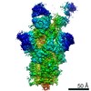
Journal: Science / Year: 2020 Title: Potent neutralizing antibodies from COVID-19 patients define multiple targets of vulnerability. Authors: Philip J M Brouwer / Tom G Caniels / Karlijn van der Straten / Jonne L Snitselaar / Yoann Aldon / Sandhya Bangaru / Jonathan L Torres / Nisreen M A Okba / Mathieu Claireaux / Gius Kerster / ...Authors: Philip J M Brouwer / Tom G Caniels / Karlijn van der Straten / Jonne L Snitselaar / Yoann Aldon / Sandhya Bangaru / Jonathan L Torres / Nisreen M A Okba / Mathieu Claireaux / Gius Kerster / Arthur E H Bentlage / Marlies M van Haaren / Denise Guerra / Judith A Burger / Edith E Schermer / Kirsten D Verheul / Niels van der Velde / Alex van der Kooi / Jelle van Schooten / Mariëlle J van Breemen / Tom P L Bijl / Kwinten Sliepen / Aafke Aartse / Ronald Derking / Ilja Bontjer / Neeltje A Kootstra / W Joost Wiersinga / Gestur Vidarsson / Bart L Haagmans / Andrew B Ward / Godelieve J de Bree / Rogier W Sanders / Marit J van Gils / Abstract: The rapid spread of severe acute respiratory syndrome coronavirus 2 (SARS-CoV-2) has had a large impact on global health, travel, and economy. Therefore, preventative and therapeutic measures are ...The rapid spread of severe acute respiratory syndrome coronavirus 2 (SARS-CoV-2) has had a large impact on global health, travel, and economy. Therefore, preventative and therapeutic measures are urgently needed. Here, we isolated monoclonal antibodies from three convalescent coronavirus disease 2019 (COVID-19) patients using a SARS-CoV-2 stabilized prefusion spike protein. These antibodies had low levels of somatic hypermutation and showed a strong enrichment in VH1-69, VH3-30-3, and VH1-24 gene usage. A subset of the antibodies was able to potently inhibit authentic SARS-CoV-2 infection at a concentration as low as 0.007 micrograms per milliliter. Competition and electron microscopy studies illustrate that the SARS-CoV-2 spike protein contains multiple distinct antigenic sites, including several receptor-binding domain (RBD) epitopes as well as non-RBD epitopes. In addition to providing guidance for vaccine design, the antibodies described here are promising candidates for COVID-19 treatment and prevention.
History
Deposition
May 27, 2020
-
Header (metadata) release
Jul 1, 2020
-
Map release
Jul 1, 2020
-
Update
Aug 19, 2020
-
Current status
Aug 19, 2020
Processing site: RCSB / Status: Released
-
Structure visualization
Movie
Surface view with section colored by density value
In the structure databanks used in Yorodumi, some data are registered as the other names, "COVID-19 virus" and "2019-nCoV". Here are the details of the virus and the list of structure data.
Jan 31, 2019. EMDB accession codes are about to change! (news from PDBe EMDB page)
EMDB accession codes are about to change! (news from PDBe EMDB page)
The allocation of 4 digits for EMDB accession codes will soon come to an end. Whilst these codes will remain in use, new EMDB accession codes will include an additional digit and will expand incrementally as the available range of codes is exhausted. The current 4-digit format prefixed with “EMD-” (i.e. EMD-XXXX) will advance to a 5-digit format (i.e. EMD-XXXXX), and so on. It is currently estimated that the 4-digit codes will be depleted around Spring 2019, at which point the 5-digit format will come into force.
The EM Navigator/Yorodumi systems omit the EMD- prefix.
Related info.:Q: What is EMD? / ID/Accession-code notation in Yorodumi/EM Navigator
Yorodumi is a browser for structure data from EMDB, PDB, SASBDB, etc.
This page is also the successor to EM Navigator detail page, and also detail information page/front-end page for Omokage search.
The word "yorodu" (or yorozu) is an old Japanese word meaning "ten thousand". "mi" (miru) is to see.
Related info.:EMDB / PDB / SASBDB / Comparison of 3 databanks / Yorodumi Search / Aug 31, 2016. New EM Navigator & Yorodumi / Yorodumi Papers / Jmol/JSmol / Function and homology information / Changes in new EM Navigator and Yorodumi
 Movie
Movie Controller

 Yorodumi
 Open data
 Basic information
 Map data
 Sample
 Function and homology information
 Homo sapiens (human)
 Authors
 United States, 1 items
  Citation
 Structure visualization
 UCSF Chimera
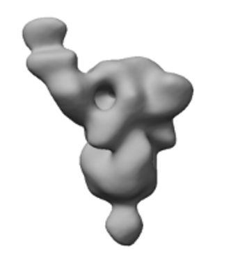
 Downloads & links
 emd_22062.png
 http://ftp.pdbj.org/pub/emdb/structures/EMD-22062
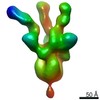


 Z (Sec.)
 Y (Row.)
 X (Col.)


 Sample components
 Processing
 Electron microscopy
