 Movie
Movie Controller

[English] 日本語
 Yorodumi
- EMDB-2101: Electron tomography of forming Hepatitis C virus-like particles i... -
+ Open data

- Basic information
| Entry | Database: EMDB / ID: EMD-2101 | |||||||||
|---|---|---|---|---|---|---|---|---|---|---|
| Title | Electron tomography of forming Hepatitis C virus-like particles in insect cells. | |||||||||
 Map data Map data | Tomographic reconstruction of an insect cell Sf9 producing Hepatitis C virus-like particles. | |||||||||
 Sample Sample |
| |||||||||
 Keywords Keywords | Hepatitis C virus / Hepatitis C virus-like particles / virus morphogenesis / budding | |||||||||
| Biological species | Hepatitis C Virus-like particles | |||||||||
| Method | electron tomography / negative staining | |||||||||
 Authors Authors | Badia-Martinez D / Peralta B / Andres G / Guerra M / Gil-Carton D / Abrescia NG | |||||||||
 Citation Citation | Journal: Virology / Year: 2012 Title: Three-dimensional visualization of forming Hepatitis C virus-like particles by electron-tomography. Authors: Daniel Badia-Martinez / Bibiana Peralta / German Andrés / Milagros Guerra / David Gil-Carton / Nicola G A Abrescia /  Abstract: Hepatitis C virus infects almost 170 million people per year but its assembly pathway, architecture and the structures of its envelope proteins are poorly understood. Using electron tomography of ...Hepatitis C virus infects almost 170 million people per year but its assembly pathway, architecture and the structures of its envelope proteins are poorly understood. Using electron tomography of plastic-embedded sections of insect cells, we have visualized the morphogenesis of recombinant Hepatitis C virus-like particles. Our data provide a three-dimensional sketch of viral assembly at the endoplasmic reticulum showing different budding stages and contiguity of buds. This latter phenomenon could play an important role during the assembly of wt-HCV and explain the size-heterogeneity of its particles. | |||||||||
| History |
|
- Structure visualization
| Movie |
 Movie viewer Movie viewer |
|---|---|
| Structure viewer | EM map: SurfViewMolmilJmol/JSmol |
| Supplemental images |

- Downloads & links
-EMDB archive
| Map data | emd_2101.map.gz | 1.7 GB | EMDB map data format | |
|---|---|---|---|---|
| Header (meta data) | emd-2101-v30.xmlemd-2101.xml | 8.6 KB 8.6 KB | Display Display | EMDB header |
| Images | 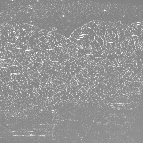 EMD-2101.png EMD-2101.png | 289.2 KB | ||
| Archive directory |  http://ftp.pdbj.org/pub/emdb/structures/EMD-2101ftp://ftp.pdbj.org/pub/emdb/structures/EMD-2101 http://ftp.pdbj.org/pub/emdb/structures/EMD-2101ftp://ftp.pdbj.org/pub/emdb/structures/EMD-2101 | HTTPS FTP |
-Validation report
| Summary document | emd_2101_validation.pdf.gz | 144.2 KB | Display | EMDB validaton report |
|---|---|---|---|---|
| Full document | emd_2101_full_validation.pdf.gz | 143.3 KB | Display | |
| Data in XML | emd_2101_validation.xml.gz | 4.9 KB | Display | |
| Arichive directory | https://ftp.pdbj.org/pub/emdb/validation_reports/EMD-2101ftp://ftp.pdbj.org/pub/emdb/validation_reports/EMD-2101 | HTTPS FTP |
-Links
| EMDB pages | EMDB (EBI/PDBe) / EMDataResource |
|---|
-Map
| File | Download / File: emd_2101.map.gz / Format: CCP4 / Size: 1.9 GB / Type: IMAGE STORED AS FLOATING POINT NUMBER (4 BYTES) | ||||||||||||||||||||||||||||||||||||||||||||||||||||||||||||||||||||
|---|---|---|---|---|---|---|---|---|---|---|---|---|---|---|---|---|---|---|---|---|---|---|---|---|---|---|---|---|---|---|---|---|---|---|---|---|---|---|---|---|---|---|---|---|---|---|---|---|---|---|---|---|---|---|---|---|---|---|---|---|---|---|---|---|---|---|---|---|---|
| Annotation | Tomographic reconstruction of an insect cell Sf9 producing Hepatitis C virus-like particles. | ||||||||||||||||||||||||||||||||||||||||||||||||||||||||||||||||||||
| Voxel size | X=Y=Z: 9.46 Å | ||||||||||||||||||||||||||||||||||||||||||||||||||||||||||||||||||||
| Density |
| ||||||||||||||||||||||||||||||||||||||||||||||||||||||||||||||||||||
| Symmetry | Space group: 1 | ||||||||||||||||||||||||||||||||||||||||||||||||||||||||||||||||||||
| Details | EMDB XML:
CCP4 map header:
| ||||||||||||||||||||||||||||||||||||||||||||||||||||||||||||||||||||
-Supplemental data
- Sample components
-Entire : Tomographic reconstruction of an insect cell Sf9 producing Hepati...
| Entire | Name: Tomographic reconstruction of an insect cell Sf9 producing Hepatitis C virus-like particles |
|---|---|
| Components |
|
-Supramolecule #1000: Tomographic reconstruction of an insect cell Sf9 producing Hepati...
| Supramolecule | Name: Tomographic reconstruction of an insect cell Sf9 producing Hepatitis C virus-like particles type: sample / ID: 1000 / Number unique components: 1 |
|---|
-Supramolecule #1: Hepatitis C virus-like particles
| Supramolecule | Name: Hepatitis C virus-like particles / type: organelle_or_cellular_component / ID: 1 / Name.synonym: HCV-LP / Recombinant expression: No / Database: NCBI |
|---|---|
| Source (natural) | Organism: Hepatitis C Virus-like particles / Cell: Sf9 insect cells / Location in cell: Endoplasmic reticulum |
-Experimental details
-Structure determination
| Method | negative staining |
|---|---|
 Processing Processing | electron tomography |
-Sample preparation
| Staining | Type: NEGATIVE Details: cells were fixed with 2% glutaraldehyde in PBS (pH 7.4), stained with 1% OsO4 and processed for conventional Epon resin embedding |
|---|---|
| Grid | Details: 200 mesh copper EM grids counterstained with uranyl acetate and lead citrate |
| Vitrification | Cryogen name: NONE / Instrument: OTHER |
- Electron microscopy
| Microscope | JEOL 2200FS |
|---|---|
| Temperature | Min: 292 K / Max: 294 K / Average: 293 K |
| Specialist optics | Energy filter - Name: omega |
| Date | Nov 11, 2011 |
| Image recording | Category: CCD / Film or detector model: TVIPS TEMCAM-F416 (4k x 4k) / Number real images: 126 / Bits/pixel: 32 |
| Electron beam | Acceleration voltage: 200 kV / Electron source:  FIELD EMISSION GUN FIELD EMISSION GUN |
| Electron optics | Illumination mode: FLOOD BEAM / Imaging mode: BRIGHT FIELD / Nominal defocus max: -7.0 µm / Nominal defocus min: -5.0 µm / Nominal magnification: 25000 |
| Sample stage | Specimen holder model: JEOL / Tilt series - Axis1 - Min angle: -65 ° / Tilt series - Axis1 - Max angle: 65 ° / Tilt series - Axis1 - Angle increment: 1.5 ° |
-Image processing
| Final reconstruction | Algorithm: OTHER / Software - Name: IMOD, TOMO3D / Number images used: 126 |
|---|
