 Movie
Movie Controller


+ Open data

- Basic information
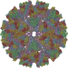
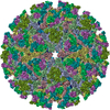
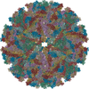
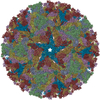
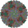
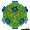
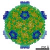
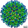
| Entry | Database: PDB / ID: 7ko8 | |||||||||||||||
|---|---|---|---|---|---|---|---|---|---|---|---|---|---|---|---|---|
| Title | Cryo-EM structure of the mature and infective Mayaro virus | |||||||||||||||
 Components Components |
| |||||||||||||||
 Keywords Keywords | VIRUS / alphavirus / mayaro virus / single particle cryo-EM / mature and infective viral particle | |||||||||||||||
| Biological species |  Mayaro virus Mayaro virus | |||||||||||||||
| Method | ELECTRON MICROSCOPY / single particle reconstruction / cryo EM / Resolution: 4.4 Å | |||||||||||||||
 Authors Authors | Riberio-Filho, H.V. / Coimbra, L.D. / Cassago, A. / Rocha, R.P.F. / Padilha, A.C.M. / Schatz, M. / van Heel, M.G. / Portugal, R.V. / Trivella, D.B.B. / de Oliveira, P.S.L. / Marques, R.E. | |||||||||||||||
| Funding support |  Brazil, 4items Brazil, 4items
| |||||||||||||||
 Citation Citation | Journal: Nat Commun / Year: 2021 Title: Cryo-EM structure of the mature and infective Mayaro virus at 4.4 Å resolution reveals features of arthritogenic alphaviruses. Authors: Helder V Ribeiro-Filho / Lais D Coimbra / Alexandre Cassago / Rebeca P F Rocha / João Victor da Silva Guerra / Rafael de Felicio / Carolina Moretto Carnieli / Luiza Leme / Antonio Cláudio ...Authors: Helder V Ribeiro-Filho / Lais D Coimbra / Alexandre Cassago / Rebeca P F Rocha / João Victor da Silva Guerra / Rafael de Felicio / Carolina Moretto Carnieli / Luiza Leme / Antonio Cláudio Padilha / Adriana F Paes Leme / Daniela B B Trivella / Rodrigo Villares Portugal / Paulo Sérgio Lopes-de-Oliveira / Rafael Elias Marques / Abstract: Mayaro virus (MAYV) is an emerging arbovirus of the Americas that may cause a debilitating arthritogenic disease. The biology of MAYV is not fully understood and largely inferred from related ...Mayaro virus (MAYV) is an emerging arbovirus of the Americas that may cause a debilitating arthritogenic disease. The biology of MAYV is not fully understood and largely inferred from related arthritogenic alphaviruses. Here, we present the structure of MAYV at 4.4 Å resolution, obtained from a preparation of mature, infective virions. MAYV presents typical alphavirus features and organization. Interactions between viral proteins that lead to particle formation are described together with a hydrophobic pocket formed between E1 and E2 spike proteins and conformational epitopes specific of MAYV. We also describe MAYV glycosylation residues in E1 and E2 that may affect MXRA8 host receptor binding, and a molecular "handshake" between MAYV spikes formed by N262 glycosylation in adjacent E2 proteins. The structure of MAYV is suggestive of structural and functional complexity among alphaviruses, which may be targeted for specificity or antiviral activity. | |||||||||||||||
| History |
|
- Structure visualization
| Movie |
 Movie viewer Movie viewer |
|---|---|
| Structure viewer | Molecule: MolmilJmol/JSmol |
- Downloads & links
-Download
| PDBx/mmCIF format | 7ko8.cif.gz | 680.3 KB | Display | PDBx/mmCIF format |
|---|---|---|---|---|
| PDB format | pdb7ko8.ent.gz | 540.5 KB | Display | PDB format |
| PDBx/mmJSON format | 7ko8.json.gz | Tree view | PDBx/mmJSON format | |
| Others |  Other downloads Other downloads |
-Validation report
| Arichive directory | https://data.pdbj.org/pub/pdb/validation_reports/ko/7ko8ftp://data.pdbj.org/pub/pdb/validation_reports/ko/7ko8 | HTTPS FTP |
|---|
-Related structure data
| Related structure data | 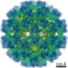 22961MC M: map data used to model this data C: citing same article ( |
|---|---|
| Similar structure data |

-Links
 PDBj
PDBj- Assembly
| Deposited unit | 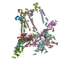
|
|---|---|
| 1 | x 60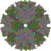
|
| 2 |
|
| 3 | x 5
|
| 4 | x 6
|
| 5 | 
|
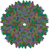
| Symmetry | Point symmetry: (Schoenflies symbol: I (icosahedral)) |
-Components
-Protein , 3 types, 12 molecules ADGJBEHLCFIM
| #1: Protein | Mass: 29399.160 Da / Num. of mol.: 4 / Source method: isolated from a natural source / Source: (natural) Mayaro virus / Strain: IQT4235#2: Protein | Mass: 47339.621 Da / Num. of mol.: 4 / Source method: isolated from a natural source / Source: (natural) Mayaro virus / Strain: IQT4235#3: Protein | Mass: 46646.066 Da / Num. of mol.: 4 / Source method: isolated from a natural source / Source: (natural) Mayaro virus / Strain: IQT4235 |
|---|
-Sugars , 3 types, 8 molecules
| #4: Polysaccharide | Source method: isolated from a genetically manipulated source #5: Polysaccharide | alpha-D-mannopyranose-(1-3)-[alpha-D-mannopyranose-(1-6)]beta-D-mannopyranose-(1-4)-2-acetamido-2- ...alpha-D-mannopyranose-(1-3)-[alpha-D-mannopyranose-(1-6)]beta-D-mannopyranose-(1-4)-2-acetamido-2-deoxy-beta-D-glucopyranose-(1-4)-2-acetamido-2-deoxy-beta-D-glucopyranose Source method: isolated from a genetically manipulated source #6: Polysaccharide | 2-acetamido-2-deoxy-beta-D-glucopyranose-(1-4)-2-acetamido-2-deoxy-beta-D-glucopyranose | Source method: isolated from a genetically manipulated source |
|---|
-Details
| Has ligand of interest | Y |
|---|---|
| Has protein modification | Y |
-Experimental details
-Experiment
| Experiment | Method: ELECTRON MICROSCOPY |
|---|---|
| EM experiment | Aggregation state: PARTICLE / 3D reconstruction method: single particle reconstruction |
- Sample preparation
| Component | Name: Mayaro virus / Type: VIRUS / Details: Mayaro virus was purified from Vero CCL81 cells. / Entity ID: #1-#3 / Source: NATURAL |
|---|---|
| Source (natural) | Organism: Mayaro virus |
| Details of virus | Empty: NO / Enveloped: YES / Isolate: STRAIN / Type: VIRION |
| Buffer solution | pH: 7 |
| Specimen | Embedding applied: NO / Shadowing applied: NO / Staining applied: NO / Vitrification applied: YES |
| Vitrification | Cryogen name: ETHANE |
- Electron microscopy imaging
| Experimental equipment |  Model: Titan Krios / Image courtesy: FEI Company |
|---|---|
| Microscopy | Model: FEI TITAN KRIOS |
| Electron gun | Electron source:  FIELD EMISSION GUN / Accelerating voltage: 300 kV / Illumination mode: OTHER FIELD EMISSION GUN / Accelerating voltage: 300 kV / Illumination mode: OTHER |
| Electron lens | Mode: BRIGHT FIELD |
| Image recording | Electron dose: 30 e/Å2 / Film or detector model: FEI FALCON III (4k x 4k) |
- Processing
| Software | Name: PHENIX / Version: 1.16_3549: / Classification: refinement | ||||||||||||||||||||||||
|---|---|---|---|---|---|---|---|---|---|---|---|---|---|---|---|---|---|---|---|---|---|---|---|---|---|
| EM software | Name: IMAGIC / Category: 3D reconstruction | ||||||||||||||||||||||||
| CTF correction | Type: PHASE FLIPPING ONLY | ||||||||||||||||||||||||
| Symmetry | Point symmetry: I (icosahedral) | ||||||||||||||||||||||||
| 3D reconstruction | Resolution: 4.4 Å / Resolution method: FSC 1/2 BIT CUT-OFF / Num. of particles: 40179 / Symmetry type: POINT | ||||||||||||||||||||||||
| Atomic model building | Protocol: FLEXIBLE FIT / Space: REAL | ||||||||||||||||||||||||
| Refine LS restraints |
|
