Movie
Movie Controller


+ Open data

- Basic information
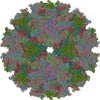
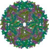
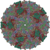
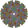
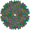
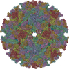
| Entry | Database: PDB / ID: 3j2w | ||||||
|---|---|---|---|---|---|---|---|
| Title | Electron cryo-microscopy of Chikungunya virus | ||||||
 Components Components |
| ||||||
 Keywords Keywords | VIRUS / E1-E2 glycoprotein / nucleocapsid protein / transmembrane helix | ||||||
| Function / homology |  Function and homology information Function and homology informationtogavirin / T=4 icosahedral viral capsid / host cell endoplasmic reticulum / channel activity / monoatomic ion transmembrane transport / symbiont-mediated suppression of host toll-like receptor signaling pathway / host cell Golgi apparatus / entry receptor-mediated virion attachment to host cell / serine-type endopeptidase activity / fusion of virus membrane with host endosome membrane ...togavirin / T=4 icosahedral viral capsid / host cell endoplasmic reticulum / channel activity / monoatomic ion transmembrane transport / symbiont-mediated suppression of host toll-like receptor signaling pathway / host cell Golgi apparatus / entry receptor-mediated virion attachment to host cell / serine-type endopeptidase activity / fusion of virus membrane with host endosome membrane / symbiont entry into host cell / host cell nucleus / host cell plasma membrane / virion membrane / structural molecule activity / proteolysis / RNA binding / identical protein binding Similarity search - Function | ||||||
| Biological species |   Chikungunya virus Chikungunya virus | ||||||
| Method | ELECTRON MICROSCOPY / single particle reconstruction / cryo EM / Resolution: 5 Å | ||||||
 Authors Authors | Sun, S. / Xiang, Y. / Rossmann, M.G. | ||||||
 Citation Citation | Journal: Elife / Year: 2013 Title: Structural analyses at pseudo atomic resolution of Chikungunya virus and antibodies show mechanisms of neutralization. Authors: Siyang Sun / Ye Xiang / Wataru Akahata / Heather Holdaway / Pankaj Pal / Xinzheng Zhang / Michael S Diamond / Gary J Nabel / Michael G Rossmann /  Abstract: A 5.3 Å resolution, cryo-electron microscopy (cryoEM) map of Chikungunya virus-like particles (VLPs) has been interpreted using the previously published crystal structure of the Chikungunya E1-E2 ...A 5.3 Å resolution, cryo-electron microscopy (cryoEM) map of Chikungunya virus-like particles (VLPs) has been interpreted using the previously published crystal structure of the Chikungunya E1-E2 glycoprotein heterodimer. The heterodimer structure was divided into domains to obtain a good fit to the cryoEM density. Differences in the T = 4 quasi-equivalent heterodimer components show their adaptation to different environments. The spikes on the icosahedral 3-fold axes and those in general positions are significantly different, possibly representing different phases during initial generation of fusogenic E1 trimers. CryoEM maps of neutralizing Fab fragments complexed with VLPs have been interpreted using the crystal structures of the Fab fragments and the VLP structure. Based on these analyses the CHK-152 antibody was shown to stabilize the viral surface, hindering the exposure of the fusion-loop, likely neutralizing infection by blocking fusion. The CHK-9, m10 and m242 antibodies surround the receptor-attachment site, probably inhibiting infection by blocking cell attachment. DOI:http://dx.doi.org/10.7554/eLife.00435.001. | ||||||
| History |
|
- Structure visualization
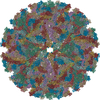
| Movie |
Movie viewer |
|---|---|
| Structure viewer | Molecule: MolmilJmol/JSmol |
- Downloads & links
-Download
| PDBx/mmCIF format | 3j2w.cif.gz | 775.1 KB | Display | PDBx/mmCIF format |
|---|---|---|---|---|
| PDB format | pdb3j2w.ent.gz | 621.5 KB | Display | PDB format |
| PDBx/mmJSON format | 3j2w.json.gz | Tree view | PDBx/mmJSON format | |
| Others |  Other downloads Other downloads |
-Validation report
| Arichive directory | https://data.pdbj.org/pub/pdb/validation_reports/j2/3j2wftp://data.pdbj.org/pub/pdb/validation_reports/j2/3j2w | HTTPS FTP |
|---|
-Related structure data
| Related structure data | 5577MC 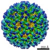 5576C 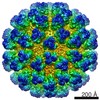 5578C  5579C  5580C  3j2xC  3j2yC  3j2zC  3j30C  4gq9C  4gq8 M: map data used to model this data C: citing same article ( |
|---|---|
| Similar structure data |

-Links
 PDBj
PDBj


- Assembly
| Deposited unit | 
|
|---|---|
| 1 | x 60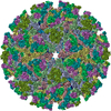
|
| 2 |
|
| 3 | x 5
|
| 4 | x 6
|
| 5 | 
|
| Symmetry | Point symmetry: (Schoenflies symbol: I (icosahedral)) |
-Components
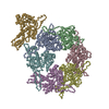
-Glycoprotein ... , 5 types, 16 molecules ABCDMNOPEFGHQRST
| #1: Protein | Mass: 42712.309 Da / Num. of mol.: 4 / Fragment: UNP residues 810-1202 Source method: isolated from a genetically manipulated source Source: (gene. exp.) Chikungunya virus / Strain: 37997 / Cell line (production host): HEK 293F / Production host:  Homo sapiens (human) / References: UniProt: Q1H8W5 Homo sapiens (human) / References: UniProt: Q1H8W5#2: Protein | Mass: 37848.852 Da / Num. of mol.: 3 / Fragment: UNP residues 332-667 Source method: isolated from a genetically manipulated source Source: (gene. exp.) Chikungunya virus / Strain: 37997 / Cell line (production host): HEK 293F / Production host: Homo sapiens (human) / References: UniProt: Q1H8W5#3: Protein | | Mass: 37866.891 Da / Num. of mol.: 1 / Fragment: UNP residues 332-667 Source method: isolated from a genetically manipulated source Source: (gene. exp.) Chikungunya virus / Strain: 37997 / Cell line (production host): HEK 293F / Production host: Homo sapiens (human) / References: UniProt: Q1H8W5#4: Protein/peptide | Mass: 4810.723 Da / Num. of mol.: 4 / Fragment: UNP residues 668-748 Source method: isolated from a genetically manipulated source Source: (gene. exp.) Chikungunya virus / Strain: 37997 / Cell line (production host): HEK 293F / Production host: Homo sapiens (human) / References: UniProt: Q5XXP3#5: Protein | Mass: 8817.458 Da / Num. of mol.: 4 / Fragment: UNP residues 1203-1248 Source method: isolated from a genetically manipulated source Source: (gene. exp.) Chikungunya virus / Strain: 37997 / Cell line (production host): HEK 293F / Production host: Homo sapiens (human) / References: UniProt: Q5XXP3 |
|---|
-Protein , 1 types, 4 molecules IJKL
| #6: Protein | Mass: 16229.508 Da / Num. of mol.: 4 / Fragment: UNP residues 113-261 Source method: isolated from a genetically manipulated source Source: (gene. exp.) Chikungunya virus / Strain: 37997 / Cell line (production host): HEK 293F / Production host: Homo sapiens (human) / References: UniProt: Q5XXP3 |
|---|
-Details
| Has protein modification | Y |
|---|
-Experimental details
-Experiment
| Experiment | Method: ELECTRON MICROSCOPY |
|---|---|
| EM experiment | Aggregation state: PARTICLE / 3D reconstruction method: single particle reconstruction |
- Sample preparation
| Component | Name: Chikungunya VLP / Type: VIRUS |
|---|---|
| Details of virus | Empty: NO / Enveloped: YES / Host category: INVERTEBRATES / Isolate: STRAIN / Type: VIRUS-LIKE PARTICLE |
| Natural host | Organism: Aedes albopictus |
| Buffer solution | Name: PBS / pH: 7 / Details: PBS |
| Specimen | Embedding applied: NO / Shadowing applied: NO / Staining applied: NO / Vitrification applied: YES |
| Specimen support | Details: 400 mesh copper grid (1.2 um hole) |
| Vitrification | Instrument: FEI VITROBOT MARK I / Cryogen name: ETHANE / Temp: 100 K / Humidity: 100 % Details: Blot for 2 seconds before plunging into liquid ethane (FEI VITROBOT MARK I) Method: Blot for 2 seconds before plunging |
- Electron microscopy imaging
| Experimental equipment |  Model: Titan Krios / Image courtesy: FEI Company |
|---|---|
| Microscopy | Model: FEI TITAN KRIOS / Date: Jan 1, 2011 |
| Electron gun | Electron source:  FIELD EMISSION GUN / Accelerating voltage: 300 kV / Illumination mode: FLOOD BEAM FIELD EMISSION GUN / Accelerating voltage: 300 kV / Illumination mode: FLOOD BEAM |
| Electron lens | Mode: BRIGHT FIELD / Nominal magnification: 59000 X / Cs: 2.7 mm Astigmatism: Objective lens astigmatism was corrected at 250000 times magnification Camera length: 0 mm |
| Specimen holder | Specimen holder model: FEI TITAN KRIOS AUTOGRID HOLDER / Tilt angle max: 0 ° / Tilt angle min: 0 ° |
| Image recording | Electron dose: 25 e/Å2 / Film or detector model: GENERIC FILM |
| Image scans | Num. digital images: 1532 |
| Radiation | Protocol: SINGLE WAVELENGTH / Monochromatic (M) / Laue (L): M / Scattering type: x-ray |
| Radiation wavelength | Relative weight: 1 |
- Processing
| EM software |
| ||||||||||||
|---|---|---|---|---|---|---|---|---|---|---|---|---|---|
| CTF correction | Details: each particle | ||||||||||||
| Symmetry | Point symmetry: I (icosahedral) | ||||||||||||
| 3D reconstruction | Resolution: 5 Å / Resolution method: FSC 0.5 CUT-OFF / Num. of particles: 36236 / Details: (Single particle--Applied symmetry: I) / Symmetry type: POINT | ||||||||||||
| Atomic model building | Protocol: RIGID BODY FIT / Space: RECIPROCAL / Details: REFINEMENT PROTOCOL--rigid body | ||||||||||||
| Atomic model building | PDB-ID: 3N43 Accession code: 3N43 / Source name: PDB / Type: experimental model | ||||||||||||
| Refinement step | Cycle: LAST
|