Movie
Movie Controller

[English] 日本語
 Yorodumi
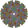
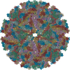
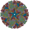
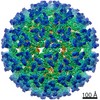
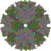
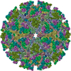
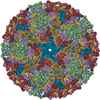
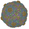
- PDB-6nk6: Electron Cryo-Microscopy Of Chikungunya VLP in complex with mouse... -
+ Open data

- Basic information
| Entry | Database: PDB / ID: 6nk6 | ||||||
|---|---|---|---|---|---|---|---|
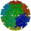
| Title | Electron Cryo-Microscopy Of Chikungunya VLP in complex with mouse Mxra8 receptor | ||||||
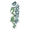
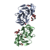
 Components Components |
| ||||||
 Keywords Keywords | VIRUS LIKE PARTICLE/SIGNALING PROTEIN / Chikungunya / virus-like particle / viral receptor / Mxra8 / Structural Genomics / Center for Structural Genomics of Infectious Diseases / CSGID / VIRUS LIKE PARTICLE-SIGNALING PROTEIN complex | ||||||
| Function / homology |  Function and homology information Function and homology informationestablishment of glial blood-brain barrier / Regulation of Insulin-like Growth Factor (IGF) transport and uptake by Insulin-like Growth Factor Binding Proteins (IGFBPs) / Post-translational protein phosphorylation / togavirin / T=4 icosahedral viral capsid / ciliary membrane / host cell endoplasmic reticulum / bicellular tight junction / channel activity / monoatomic ion transmembrane transport ...establishment of glial blood-brain barrier / Regulation of Insulin-like Growth Factor (IGF) transport and uptake by Insulin-like Growth Factor Binding Proteins (IGFBPs) / Post-translational protein phosphorylation / togavirin / T=4 icosahedral viral capsid / ciliary membrane / host cell endoplasmic reticulum / bicellular tight junction / channel activity / monoatomic ion transmembrane transport / symbiont-mediated suppression of host toll-like receptor signaling pathway / host cell Golgi apparatus / entry receptor-mediated virion attachment to host cell / cell differentiation / cell adhesion / serine-type endopeptidase activity / fusion of virus membrane with host endosome membrane / symbiont entry into host cell / host cell nucleus / host cell plasma membrane / virion membrane / structural molecule activity / cell surface / proteolysis / RNA binding / nucleus / cytoplasm Similarity search - Function | ||||||
| Biological species |   Chikungunya virus strain Senegal 37997 Chikungunya virus strain Senegal 37997 | ||||||
| Method | ELECTRON MICROSCOPY / single particle reconstruction / cryo EM / Resolution: 4.06 Å | ||||||
 Authors Authors | Basore, K. / Kim, A.S. / Nelson, C.A. / Fremont, D.H. / Center for Structural Genomics of Infectious Diseases (CSGID) | ||||||
| Funding support |  United States, 1items United States, 1items
| ||||||
 Citation Citation | Journal: Cell / Year: 2019 Title: Cryo-EM Structure of Chikungunya Virus in Complex with the Mxra8 Receptor. Authors: Katherine Basore / Arthur S Kim / Christopher A Nelson / Rong Zhang / Brittany K Smith / Carla Uranga / Lo Vang / Ming Cheng / Michael L Gross / Jonathan Smith / Michael S Diamond / Daved H Fremont / Abstract: Mxra8 is a receptor for multiple arthritogenic alphaviruses that cause debilitating acute and chronic musculoskeletal disease in humans. Herein, we present a 2.2 Å resolution X-ray crystal ...Mxra8 is a receptor for multiple arthritogenic alphaviruses that cause debilitating acute and chronic musculoskeletal disease in humans. Herein, we present a 2.2 Å resolution X-ray crystal structure of Mxra8 and 4 to 5 Å resolution cryo-electron microscopy reconstructions of Mxra8 bound to chikungunya (CHIKV) virus-like particles and infectious virus. The Mxra8 ectodomain contains two strand-swapped Ig-like domains oriented in a unique disulfide-linked head-to-head arrangement. Mxra8 binds by wedging into a cleft created by two adjacent CHIKV E2-E1 heterodimers in one trimeric spike and engaging a neighboring spike. Two binding modes are observed with the fully mature VLP, with one Mxra8 binding with unique contacts. Only the high-affinity binding mode was observed in the complex with infectious CHIKV, as viral maturation and E3 occupancy appear to influence receptor binding-site usage. Our studies provide insight into how Mxra8 binds CHIKV and creates a path for developing alphavirus entry inhibitors. | ||||||
| History |
|
- Structure visualization
| Movie |
Movie viewer |
|---|---|
| Structure viewer | Molecule: MolmilJmol/JSmol |
 UCSF Chimera
- Downloads & links
-Download
| PDBx/mmCIF format | 6nk6.cif.gz | 905.7 KB | Display | PDBx/mmCIF format |
|---|---|---|---|---|
| PDB format | pdb6nk6.ent.gz | 745.7 KB | Display | PDB format |
| PDBx/mmJSON format | 6nk6.json.gz | Tree view | PDBx/mmJSON format | |
| Others |  Other downloads Other downloads |
-Validation report
| Arichive directory | https://data.pdbj.org/pub/pdb/validation_reports/nk/6nk6ftp://data.pdbj.org/pub/pdb/validation_reports/nk/6nk6 | HTTPS FTP |
|---|
-Related structure data
| Related structure data | 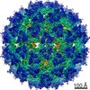 9394MC 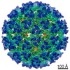 9393C 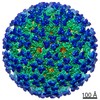 9395C 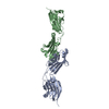 6nk3C 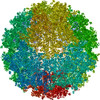 6nk5C 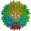 6nk7C M: map data used to model this data C: citing same article ( |
|---|---|
| Similar structure data | |
| Other databases |


-Links
 PDBj
PDBj


- Assembly
| Deposited unit | 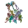
|
|---|---|
| 1 | x 60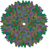
|
| 2 |
|
| 3 | x 5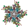
|
| 4 | x 6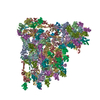
|
| 5 | 
|
| Symmetry | Point symmetry: (Schoenflies symbol: I (icosahedral)) |
-Components
| #1: Protein | Mass: 30819.457 Da / Num. of mol.: 4 / Fragment: ectodomain (UNP residues 39-291) Source method: isolated from a genetically manipulated source Source: (gene. exp.)  Homo sapiens (human) / References: UniProt: Q9DBV4 Homo sapiens (human) / References: UniProt: Q9DBV4#2: Protein | Mass: 47346.715 Da / Num. of mol.: 4 / Fragment: UNP residues 810-1248 Source method: isolated from a genetically manipulated source Source: (gene. exp.) Chikungunya virus strain Senegal 37997 / Strain: 37997 / Cell line (production host): HEK293F / Production host: Homo sapiens (human) / References: UniProt: Q5XXP3#3: Protein | Mass: 46858.312 Da / Num. of mol.: 4 / Fragment: UNP residues 330-748 Source method: isolated from a genetically manipulated source Source: (gene. exp.) Chikungunya virus strain Senegal 37997 / Strain: 37997 / Cell line (production host): HEK293F / Production host: Homo sapiens (human) / References: UniProt: Q5XXP3#4: Protein | Mass: 16458.701 Da / Num. of mol.: 4 / Fragment: UNP residues 111-261 Source method: isolated from a genetically manipulated source Source: (gene. exp.) Chikungunya virus strain Senegal 37997 / Strain: 37997 / Cell line (production host): HEK293F / Production host: Homo sapiens (human) / References: UniProt: Q5XXP3#5: Sugar | ChemComp-NAG /   Type: D-saccharide, beta linking / Mass: 221.208 Da / Num. of mol.: 12 Type: D-saccharide, beta linking / Mass: 221.208 Da / Num. of mol.: 12Source method: isolated from a genetically manipulated source Formula: C8H15NO6 Has protein modification | Y | |
|---|
-Experimental details
-Experiment
| Experiment | Method: ELECTRON MICROSCOPY |
|---|---|
| EM experiment | Aggregation state: PARTICLE / 3D reconstruction method: single particle reconstruction |
- Sample preparation
| Component |
| |||||||||||||||||||||||||||||||||||
|---|---|---|---|---|---|---|---|---|---|---|---|---|---|---|---|---|---|---|---|---|---|---|---|---|---|---|---|---|---|---|---|---|---|---|---|---|
| Molecular weight | Experimental value: NO | |||||||||||||||||||||||||||||||||||
| Source (natural) |
| |||||||||||||||||||||||||||||||||||
| Source (recombinant) |
| |||||||||||||||||||||||||||||||||||
| Details of virus | Empty: YES / Enveloped: YES / Isolate: STRAIN / Type: VIRUS-LIKE PARTICLE | |||||||||||||||||||||||||||||||||||
| Buffer solution | pH: 7.4 | |||||||||||||||||||||||||||||||||||
| Buffer component |
| |||||||||||||||||||||||||||||||||||
| Specimen | Conc.: 0.33 mg/ml / Embedding applied: NO / Shadowing applied: NO / Staining applied: NO / Vitrification applied: YES / Details: 0.33 mg/mL VLP + 15 mg/mL Mxra8 | |||||||||||||||||||||||||||||||||||
| Specimen support | Details: 0.458 mbar.l/s O2 and 0.11 mbar.l/s H2 / Grid material: COPPER / Grid mesh size: 300 divisions/in. / Grid type: Quantifoil R2/2 | |||||||||||||||||||||||||||||||||||
| Vitrification | Instrument: FEI VITROBOT MARK IV / Cryogen name: ETHANE / Humidity: 100 % / Chamber temperature: 277.15 K |
- Electron microscopy imaging
| Experimental equipment |  Model: Titan Krios / Image courtesy: FEI Company |
|---|---|
| Microscopy | Model: FEI TITAN KRIOS |
| Electron gun | Electron source: OTHER / Accelerating voltage: 300 kV / Illumination mode: FLOOD BEAM |
| Electron lens | Mode: BRIGHT FIELD |
| Image recording | Average exposure time: 0.3 sec. / Electron dose: 50 e/Å2 / Film or detector model: GATAN K2 SUMMIT (4k x 4k) |
- Processing
| EM software |
| ||||||||||||||||||||||||||||||||||||||||||
|---|---|---|---|---|---|---|---|---|---|---|---|---|---|---|---|---|---|---|---|---|---|---|---|---|---|---|---|---|---|---|---|---|---|---|---|---|---|---|---|---|---|---|---|
| CTF correction | Type: PHASE FLIPPING AND AMPLITUDE CORRECTION | ||||||||||||||||||||||||||||||||||||||||||
| Symmetry | Point symmetry: I (icosahedral) | ||||||||||||||||||||||||||||||||||||||||||
| 3D reconstruction | Resolution: 4.06 Å / Resolution method: FSC 0.143 CUT-OFF / Num. of particles: 22486 / Symmetry type: POINT | ||||||||||||||||||||||||||||||||||||||||||
| Atomic model building | Protocol: FLEXIBLE FIT / Space: REAL | ||||||||||||||||||||||||||||||||||||||||||
| Atomic model building | 3D fitting-ID: 1 / Source name: PDB / Type: experimental model
|