 Movie
Movie Controller


+ Open data

- Basic information
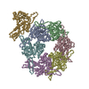
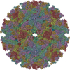
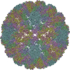
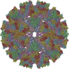
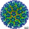
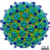
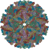
| Entry | Database: PDB / ID: 5vu2 | ||||||
|---|---|---|---|---|---|---|---|
| Title | Electron cryo-microscopy of "immature" Chikungunya VLP | ||||||
 Components Components |
| ||||||
 Keywords Keywords | VIRUS LIKE PARTICLE / Chikungunya / virus / immature | ||||||
| Function / homology |  Function and homology information Function and homology informationtogavirin / T=4 icosahedral viral capsid / host cell endoplasmic reticulum / channel activity / monoatomic ion transmembrane transport / symbiont-mediated suppression of host toll-like receptor signaling pathway / host cell Golgi apparatus / entry receptor-mediated virion attachment to host cell / serine-type endopeptidase activity / fusion of virus membrane with host endosome membrane ...togavirin / T=4 icosahedral viral capsid / host cell endoplasmic reticulum / channel activity / monoatomic ion transmembrane transport / symbiont-mediated suppression of host toll-like receptor signaling pathway / host cell Golgi apparatus / entry receptor-mediated virion attachment to host cell / serine-type endopeptidase activity / fusion of virus membrane with host endosome membrane / symbiont entry into host cell / host cell nucleus / host cell plasma membrane / virion membrane / structural molecule activity / proteolysis / RNA binding Similarity search - Function | ||||||
| Biological species |  Chikungunya virus strain Senegal 37997 Chikungunya virus strain Senegal 37997 Chikungunya virus Chikungunya virus | ||||||
| Method | ELECTRON MICROSCOPY / single particle reconstruction / cryo EM / Resolution: 6.8 Å | ||||||
 Authors Authors | Rossmann, M.G. / Yap, M.L. | ||||||
| Funding support |  United States, 1items United States, 1items
| ||||||
 Citation Citation | Journal: Proc Natl Acad Sci U S A / Year: 2017 Title: Structural studies of Chikungunya virus maturation. Authors: Moh Lan Yap / Thomas Klose / Akane Urakami / S Saif Hasan / Wataru Akahata / Michael G Rossmann /  Abstract: Cleavage of the alphavirus precursor glycoprotein p62 into the E2 and E3 glycoproteins before assembly with the nucleocapsid is the key to producing fusion-competent mature spikes on alphaviruses. ...Cleavage of the alphavirus precursor glycoprotein p62 into the E2 and E3 glycoproteins before assembly with the nucleocapsid is the key to producing fusion-competent mature spikes on alphaviruses. Here we present a cryo-EM, 6.8-Å resolution structure of an "immature" Chikungunya virus in which the cleavage site has been mutated to inhibit proteolysis. The spikes in the immature virus have a larger radius and are less compact than in the mature virus. Furthermore, domains B on the E2 glycoproteins have less freedom of movement in the immature virus, keeping the fusion loops protected under domain B. In addition, the nucleocapsid of the immature virus is more compact than in the mature virus, protecting a conserved ribosome-binding site in the capsid protein from exposure. These differences suggest that the posttranslational processing of the spikes and nucleocapsid is necessary to produce infectious virus. | ||||||
| History |
|
- Structure visualization
| Movie |
Movie viewer |
|---|---|
| Structure viewer | Molecule: MolmilJmol/JSmol |
- Downloads & links
-Download
| PDBx/mmCIF format | 5vu2.cif.gz | 874.5 KB | Display | PDBx/mmCIF format |
|---|---|---|---|---|
| PDB format | pdb5vu2.ent.gz | 671.5 KB | Display | PDB format |
| PDBx/mmJSON format | 5vu2.json.gz | Tree view | PDBx/mmJSON format | |
| Others |  Other downloads Other downloads |
-Validation report
| Arichive directory | https://data.pdbj.org/pub/pdb/validation_reports/vu/5vu2ftp://data.pdbj.org/pub/pdb/validation_reports/vu/5vu2 | HTTPS FTP |
|---|
-Related structure data
| Related structure data | 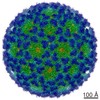 8734MC M: map data used to model this data C: citing same article ( |
|---|---|
| Similar structure data |
-Links
 PDBj
PDBj


- Assembly
| Deposited unit | 
|
|---|---|
| 1 | x 60
|
| 2 |
|
| 3 | x 5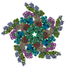
|
| 4 | x 6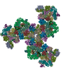
|
| 5 | 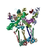
|
| Symmetry | Point symmetry: (Schoenflies symbol: I (icosahedral)) |
-Components
| #1: Protein | Mass: 95538.164 Da / Num. of mol.: 8 / Fragment: UNP residues 326-1191 Source method: isolated from a genetically manipulated source Source: (gene. exp.) Chikungunya virus strain Senegal 37997 / Strain: 37997 / Production host:  Homo sapiens (human) / References: UniProt: A0A0H4CWF2 Homo sapiens (human) / References: UniProt: A0A0H4CWF2#2: Protein | Mass: 6926.990 Da / Num. of mol.: 4 / Fragment: UNP residues 266-324 Source method: isolated from a genetically manipulated source Source: (gene. exp.) Chikungunya virus strain Senegal 37997 / Strain: 37997 / Gene: CHIKVgp2 / Production host: Homo sapiens (human) / References: UniProt: C7S7A1, UniProt: Q5XXP3*PLUS#3: Protein | Mass: 16229.508 Da / Num. of mol.: 4 / Fragment: UNP residues 113-261 Source method: isolated from a genetically manipulated source Source: (gene. exp.) Chikungunya virus (strain 37997) / Strain: 37997 / Production host: Homo sapiens (human) / References: UniProt: Q5XXP3#4: Protein/peptide | Mass: 4810.723 Da / Num. of mol.: 4 / Fragment: transmembrane helix (UNP residues 1203-1248) Source method: isolated from a genetically manipulated source Source: (gene. exp.) Chikungunya virus strain Senegal 37997 / Strain: 37997 / Production host: Homo sapiens (human) / References: UniProt: Q5XXP3#5: Protein | Mass: 8817.458 Da / Num. of mol.: 4 / Fragment: transmembrane helix (UNP residues 668-748) Source method: isolated from a genetically manipulated source Source: (gene. exp.) Chikungunya virus strain Senegal 37997 / Strain: 37997 / Production host: Homo sapiens (human) / References: UniProt: Q5XXP3Has protein modification | Y | |
|---|
-Experimental details
-Experiment
| Experiment | Method: ELECTRON MICROSCOPY |
|---|---|
| EM experiment | Aggregation state: PARTICLE / 3D reconstruction method: single particle reconstruction |
- Sample preparation
| Component | Name: Chikungunya virus strain Senegal 37997 / Type: VIRUS / Entity ID: all / Source: RECOMBINANT |
|---|---|
| Source (natural) | Organism: Chikungunya virus strain Senegal 37997 |
| Source (recombinant) | Organism: Homo sapiens (human) / Cell: Human embryonic kidney / Plasmid: pUC119 |
| Details of virus | Empty: NO / Enveloped: YES / Isolate: STRAIN / Type: VIRUS-LIKE PARTICLE |
| Buffer solution | pH: 7.4 |
| Specimen | Conc.: 3 mg/ml / Embedding applied: NO / Shadowing applied: NO / Staining applied: NO / Vitrification applied: YES |
| Vitrification | Cryogen name: ETHANE |
- Electron microscopy imaging
| Experimental equipment |  Model: Titan Krios / Image courtesy: FEI Company |
|---|---|
| Microscopy | Model: FEI TITAN KRIOS |
| Electron gun | Electron source:  FIELD EMISSION GUN / Accelerating voltage: 300 kV / Illumination mode: SPOT SCAN FIELD EMISSION GUN / Accelerating voltage: 300 kV / Illumination mode: SPOT SCAN |
| Electron lens | Mode: BRIGHT FIELD |
| Image recording | Electron dose: 36 e/Å2 / Film or detector model: GATAN K2 SUMMIT (4k x 4k) |
- Processing
| EM software |
| ||||||||||||
|---|---|---|---|---|---|---|---|---|---|---|---|---|---|
| CTF correction | Type: PHASE FLIPPING AND AMPLITUDE CORRECTION | ||||||||||||
| 3D reconstruction | Resolution: 6.8 Å / Resolution method: FSC 0.143 CUT-OFF / Num. of particles: 72944 / Symmetry type: POINT |
