 Movie
Movie Controller


+ Open data

- Basic information
| Entry | Database: PDB / ID: 6nk3 | ||||||
|---|---|---|---|---|---|---|---|
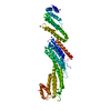
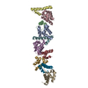
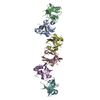
| Title | Crystal structure of murine Mxra8 ectodomain | ||||||
 Components Components | Matrix remodeling-associated protein 8 | ||||||
 Keywords Keywords | CELL INVASION / Chikungunya / virus receptor / Immunoglobulin-like / Structural Genomics / Center for Structural Genomics of Infectious Diseases / CSGID | ||||||
| Function / homology |  Function and homology information Function and homology informationestablishment of glial blood-brain barrier / Regulation of Insulin-like Growth Factor (IGF) transport and uptake by Insulin-like Growth Factor Binding Proteins (IGFBPs) / Post-translational protein phosphorylation / ciliary membrane / bicellular tight junction / cell differentiation / cell adhesion / cell surface / nucleus / cytoplasm Similarity search - Function | ||||||
| Biological species |  | ||||||
| Method |  X-RAY DIFFRACTION / SYNCHROTRON / MOLECULAR REPLACEMENT / Resolution: 2.2 Å X-RAY DIFFRACTION / SYNCHROTRON / MOLECULAR REPLACEMENT / Resolution: 2.2 Å | ||||||
 Authors Authors | Fremont, D.H. / Kim, A.S. / Nelson, C.A. / Center for Structural Genomics of Infectious Diseases (CSGID) | ||||||
| Funding support |  United States, 1items United States, 1items
| ||||||
 Citation Citation | Journal: Cell / Year: 2019 Title: Cryo-EM Structure of Chikungunya Virus in Complex with the Mxra8 Receptor. Authors: Katherine Basore / Arthur S Kim / Christopher A Nelson / Rong Zhang / Brittany K Smith / Carla Uranga / Lo Vang / Ming Cheng / Michael L Gross / Jonathan Smith / Michael S Diamond / Daved H Fremont / Abstract: Mxra8 is a receptor for multiple arthritogenic alphaviruses that cause debilitating acute and chronic musculoskeletal disease in humans. Herein, we present a 2.2 Å resolution X-ray crystal ...Mxra8 is a receptor for multiple arthritogenic alphaviruses that cause debilitating acute and chronic musculoskeletal disease in humans. Herein, we present a 2.2 Å resolution X-ray crystal structure of Mxra8 and 4 to 5 Å resolution cryo-electron microscopy reconstructions of Mxra8 bound to chikungunya (CHIKV) virus-like particles and infectious virus. The Mxra8 ectodomain contains two strand-swapped Ig-like domains oriented in a unique disulfide-linked head-to-head arrangement. Mxra8 binds by wedging into a cleft created by two adjacent CHIKV E2-E1 heterodimers in one trimeric spike and engaging a neighboring spike. Two binding modes are observed with the fully mature VLP, with one Mxra8 binding with unique contacts. Only the high-affinity binding mode was observed in the complex with infectious CHIKV, as viral maturation and E3 occupancy appear to influence receptor binding-site usage. Our studies provide insight into how Mxra8 binds CHIKV and creates a path for developing alphavirus entry inhibitors. | ||||||
| History |
|
- Structure visualization
| Structure viewer | Molecule: MolmilJmol/JSmol |
|---|
- Downloads & links
-Download
| PDBx/mmCIF format | 6nk3.cif.gz | 238.1 KB | Display | PDBx/mmCIF format |
|---|---|---|---|---|
| PDB format | pdb6nk3.ent.gz | 192.9 KB | Display | PDB format |
| PDBx/mmJSON format | 6nk3.json.gz | Tree view | PDBx/mmJSON format | |
| Others |  Other downloads Other downloads |
-Validation report
| Arichive directory | https://data.pdbj.org/pub/pdb/validation_reports/nk/6nk3ftp://data.pdbj.org/pub/pdb/validation_reports/nk/6nk3 | HTTPS FTP |
|---|
-Related structure data
| Related structure data | 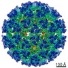 9393C 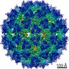 9394C 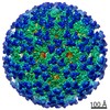 9395C 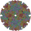 6nk5C  6nk6C  6nk7C 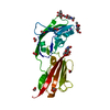 3mj6S C: citing same article ( S: Starting model for refinement |
|---|---|
| Similar structure data |


-Links
 PDBj
PDBj
- Assembly
| Deposited unit | 
| ||||||||
|---|---|---|---|---|---|---|---|---|---|
| 1 |
| ||||||||
| Unit cell |
| ||||||||
| Components on special symmetry positions |
|
-Components
| #1: Protein | Mass: 31166.775 Da / Num. of mol.: 2 / Fragment: ectodomain (UNP residues 23-296) Source method: isolated from a genetically manipulated source Source: (gene. exp.)  #2: Water | ChemComp-HOH / |  Mass: 18.015 Da / Num. of mol.: 265 / Source method: isolated from a natural source / Formula: H2O Mass: 18.015 Da / Num. of mol.: 265 / Source method: isolated from a natural source / Formula: H2OHas protein modification | Y | |
|---|
-Experimental details
-Experiment
| Experiment | Method: X-RAY DIFFRACTION / Number of used crystals: 1 |
|---|
- Sample preparation
| Crystal | Density Matthews: 4.18 Å3/Da / Density % sol: 70.56 % |
|---|---|
| Crystal grow | Temperature: 293 K / Method: vapor diffusion, hanging drop / pH: 8.5 Details: 0.1 M Tris, pH 8.5, 8% PEG8000, 25% ethylene glycol |
-Data collection
| Diffraction | Mean temperature: 293 K / Serial crystal experiment: N |
|---|---|
| Diffraction source | Source: SYNCHROTRON / Site: ALS / Beamline: 4.2.2 / Wavelength: 1.00003 Å |
| Detector | Type: RDI CMOS_8M / Detector: CMOS / Date: Oct 5, 2018 |
| Radiation | Monochromator: double crystal Si(111) / Protocol: SINGLE WAVELENGTH / Monochromatic (M) / Laue (L): M / Scattering type: x-ray |
| Radiation wavelength | Wavelength: 1.00003 Å / Relative weight: 1 |
| Reflection | Resolution: 2.2→48.46 Å / Num. obs: 51479 / % possible obs: 99.42 % / Redundancy: 26.4 % / Biso Wilson estimate: 51.15 Å2 / CC1/2: 0.999 / Net I/σ(I): 23.63 |
| Reflection shell | Resolution: 2.2→2.28 Å / Redundancy: 17.7 % / Mean I/σ(I) obs: 1.21 / Num. unique obs: 4856 / CC1/2: 0.666 / % possible all: 95.83 |
- Processing
| Software |
| ||||||||||||||||||||||||||||||||||||||||||||||||||||||||||||
|---|---|---|---|---|---|---|---|---|---|---|---|---|---|---|---|---|---|---|---|---|---|---|---|---|---|---|---|---|---|---|---|---|---|---|---|---|---|---|---|---|---|---|---|---|---|---|---|---|---|---|---|---|---|---|---|---|---|---|---|---|---|
| Refinement | Method to determine structure: MOLECULAR REPLACEMENT Starting model: PDB entry 3MJ6 Resolution: 2.2→48.46 Å / SU ML: 0.32 / Cross valid method: THROUGHOUT / σ(F): 1.34 / Phase error: 25.88
| ||||||||||||||||||||||||||||||||||||||||||||||||||||||||||||
| Solvent computation | Shrinkage radii: 0.9 Å / VDW probe radii: 1.11 Å | ||||||||||||||||||||||||||||||||||||||||||||||||||||||||||||
| Displacement parameters | Biso max: 192.52 Å2 / Biso mean: 70 Å2 / Biso min: 26.67 Å2 | ||||||||||||||||||||||||||||||||||||||||||||||||||||||||||||
| Refinement step | Cycle: final / Resolution: 2.2→48.46 Å
| ||||||||||||||||||||||||||||||||||||||||||||||||||||||||||||
| Refinement TLS params. | Method: refined / Origin x: 62.4581 Å / Origin y: 25.9824 Å / Origin z: 55.8634 Å
| ||||||||||||||||||||||||||||||||||||||||||||||||||||||||||||
| Refinement TLS group |
|
