 Movie
Movie Controller


+ Open data

- Basic information
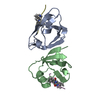
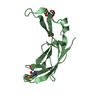
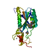
| Entry | Database: PDB / ID: 4q2o | ||||||
|---|---|---|---|---|---|---|---|
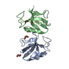
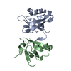
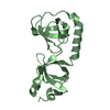
| Title | PDLIM4 PDZ in Complex with a Phage-Derived Peptide | ||||||
 Components Components | PDZ and LIM domain protein 4 | ||||||
 Keywords Keywords | PROTEIN BINDING / PDZ | ||||||
| Function / homology |  Function and homology information Function and homology informationrecycling endosome lumen / muscle structure development / excitatory chemical synaptic transmission / muscle alpha-actinin binding / filamentous actin / alpha-actinin binding / stress fiber / early endosome lumen / adherens junction / recycling endosome membrane ...recycling endosome lumen / muscle structure development / excitatory chemical synaptic transmission / muscle alpha-actinin binding / filamentous actin / alpha-actinin binding / stress fiber / early endosome lumen / adherens junction / recycling endosome membrane / Z disc / lamellipodium / actin cytoskeleton / heart development / actin binding / actin cytoskeleton organization / early endosome membrane / protein phosphatase binding / dendritic spine / cytoskeleton / postsynaptic membrane / perinuclear region of cytoplasm / protein homodimerization activity / metal ion binding / nucleus / plasma membrane / cytoplasm / cytosol Similarity search - Function | ||||||
| Biological species |  Homo sapiens (human) Homo sapiens (human) | ||||||
| Method |  X-RAY DIFFRACTION / SYNCHROTRON / MOLECULAR REPLACEMENT / Resolution: 2.1 Å X-RAY DIFFRACTION / SYNCHROTRON / MOLECULAR REPLACEMENT / Resolution: 2.1 Å | ||||||
 Authors Authors | Appleton, B.A. / Wiesmann, C. | ||||||
 Citation Citation | Journal: J.Mol.Biol. / Year: 2014 Title: A structural portrait of the PDZ domain family. Authors: Ernst, A. / Appleton, B.A. / Ivarsson, Y. / Zhang, Y. / Gfeller, D. / Wiesmann, C. / Sidhu, S.S. | ||||||
| History |
|
- Structure visualization
| Structure viewer | Molecule: MolmilJmol/JSmol |
|---|
- Downloads & links
-Download
| PDBx/mmCIF format | 4q2o.cif.gz | 212.8 KB | Display | PDBx/mmCIF format |
|---|---|---|---|---|
| PDB format | pdb4q2o.ent.gz | 173.3 KB | Display | PDB format |
| PDBx/mmJSON format | 4q2o.json.gz | Tree view | PDBx/mmJSON format | |
| Others |  Other downloads Other downloads |
-Validation report
| Arichive directory | https://data.pdbj.org/pub/pdb/validation_reports/q2/4q2oftp://data.pdbj.org/pub/pdb/validation_reports/q2/4q2o | HTTPS FTP |
|---|
-Related structure data
| Related structure data |  4q2nC  4q2pC  4q2qC 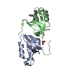 2v1wS C: citing same article ( S: Starting model for refinement |
|---|---|
| Similar structure data |


-Links
 PDBj
PDBj- Assembly
| Deposited unit | 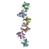
| |||||||||||||||||||||||||||||||||||||||||||||||||||||||||||||||||||||||||||||||||||||||||||||||||||||||||||||||||||||||||||||||||||||||||||||||||||||||||||||||||||||||||||||||||||||||||||||||
|---|---|---|---|---|---|---|---|---|---|---|---|---|---|---|---|---|---|---|---|---|---|---|---|---|---|---|---|---|---|---|---|---|---|---|---|---|---|---|---|---|---|---|---|---|---|---|---|---|---|---|---|---|---|---|---|---|---|---|---|---|---|---|---|---|---|---|---|---|---|---|---|---|---|---|---|---|---|---|---|---|---|---|---|---|---|---|---|---|---|---|---|---|---|---|---|---|---|---|---|---|---|---|---|---|---|---|---|---|---|---|---|---|---|---|---|---|---|---|---|---|---|---|---|---|---|---|---|---|---|---|---|---|---|---|---|---|---|---|---|---|---|---|---|---|---|---|---|---|---|---|---|---|---|---|---|---|---|---|---|---|---|---|---|---|---|---|---|---|---|---|---|---|---|---|---|---|---|---|---|---|---|---|---|---|---|---|---|---|---|---|---|---|
| 1 | 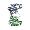
| |||||||||||||||||||||||||||||||||||||||||||||||||||||||||||||||||||||||||||||||||||||||||||||||||||||||||||||||||||||||||||||||||||||||||||||||||||||||||||||||||||||||||||||||||||||||||||||||
| 2 | 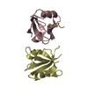
| |||||||||||||||||||||||||||||||||||||||||||||||||||||||||||||||||||||||||||||||||||||||||||||||||||||||||||||||||||||||||||||||||||||||||||||||||||||||||||||||||||||||||||||||||||||||||||||||
| 3 | 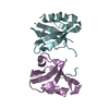
| |||||||||||||||||||||||||||||||||||||||||||||||||||||||||||||||||||||||||||||||||||||||||||||||||||||||||||||||||||||||||||||||||||||||||||||||||||||||||||||||||||||||||||||||||||||||||||||||
| Unit cell |
| |||||||||||||||||||||||||||||||||||||||||||||||||||||||||||||||||||||||||||||||||||||||||||||||||||||||||||||||||||||||||||||||||||||||||||||||||||||||||||||||||||||||||||||||||||||||||||||||
| Noncrystallographic symmetry (NCS) | NCS domain:
NCS domain segments: Refine code: 3
|