 Movie
Movie Controller

[English] 日本語
 Yorodumi
- PDB-6q0n: Structure of the Erbin PDB domain in complex with a high-affinity... -
+ Open data

- Basic information
| Entry | Database: PDB / ID: 6q0n | ||||||
|---|---|---|---|---|---|---|---|
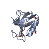
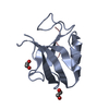
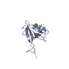
| Title | Structure of the Erbin PDB domain in complex with a high-affinity peptide | ||||||
 Components Components |
| ||||||
 Keywords Keywords | SIGNALING PROTEIN / Complex / high-affinity / specificity | ||||||
| Function / homology |  Function and homology information Function and homology informationErbB-2 class receptor binding / basal protein localization / negative regulation of nucleotide-binding oligomerization domain containing 2 signaling pathway / hemidesmosome / postsynaptic specialization / intermediate filament cytoskeleton organization / negative regulation of monocyte chemotactic protein-1 production / establishment or maintenance of epithelial cell apical/basal polarity / : / RHOB GTPase cycle ...ErbB-2 class receptor binding / basal protein localization / negative regulation of nucleotide-binding oligomerization domain containing 2 signaling pathway / hemidesmosome / postsynaptic specialization / intermediate filament cytoskeleton organization / negative regulation of monocyte chemotactic protein-1 production / establishment or maintenance of epithelial cell apical/basal polarity / : / RHOB GTPase cycle / Drug-mediated inhibition of ERBB2 signaling / Resistance of ERBB2 KD mutants to trastuzumab / Resistance of ERBB2 KD mutants to sapitinib / Resistance of ERBB2 KD mutants to tesevatinib / Resistance of ERBB2 KD mutants to neratinib / Resistance of ERBB2 KD mutants to osimertinib / Resistance of ERBB2 KD mutants to afatinib / Resistance of ERBB2 KD mutants to AEE788 / Resistance of ERBB2 KD mutants to lapatinib / Drug resistance in ERBB2 TMD/JMD mutants / RHOC GTPase cycle / response to muramyl dipeptide / basement membrane / RHOG GTPase cycle / RAC3 GTPase cycle / RHOA GTPase cycle / RAC2 GTPase cycle / regulation of postsynaptic membrane neurotransmitter receptor levels / protein targeting / Signaling by ERBB2 / RAC1 GTPase cycle / basal plasma membrane / Constitutive Signaling by Overexpressed ERBB2 / integrin-mediated signaling pathway / neuromuscular junction / Signaling by ERBB2 TMD/JMD mutants / Signaling by ERBB2 ECD mutants / Signaling by ERBB2 KD Mutants / structural constituent of cytoskeleton / cellular response to tumor necrosis factor / epidermal growth factor receptor signaling pathway / Downregulation of ERBB2 signaling / cell junction / nuclear membrane / response to lipopolysaccharide / basolateral plasma membrane / cell adhesion / nuclear speck / intracellular signal transduction / signaling receptor binding / glutamatergic synapse / signal transduction / nucleus / plasma membrane / cytoplasm Similarity search - Function | ||||||
| Biological species |  Homo sapiens (human) Homo sapiens (human)synthetic construct (others) | ||||||
| Method |  X-RAY DIFFRACTION / SYNCHROTRON / MOLECULAR REPLACEMENT / Resolution: 1.18 Å X-RAY DIFFRACTION / SYNCHROTRON / MOLECULAR REPLACEMENT / Resolution: 1.18 Å | ||||||
 Authors Authors | Singer, A.U. / Teyra, J. / Ernst, A. / Sicheri, F. / Sidhu, S.S. | ||||||
| Funding support |  Canada, 1items Canada, 1items
| ||||||
 Citation Citation | Journal: Protein Sci. / Year: 2020 Title: Comprehensive analysis of all evolutionary paths between two divergent PDZ domain specificities. Authors: Teyra, J. / Ernst, A. / Singer, A. / Sicheri, F. / Sidhu, S.S. | ||||||
| History |
|
- Structure visualization
| Structure viewer | Molecule: MolmilJmol/JSmol |
|---|
- Downloads & links
-Download
| PDBx/mmCIF format | 6q0n.cif.gz | 136.9 KB | Display | PDBx/mmCIF format |
|---|---|---|---|---|
| PDB format | pdb6q0n.ent.gz | 106.2 KB | Display | PDB format |
| PDBx/mmJSON format | 6q0n.json.gz | Tree view | PDBx/mmJSON format | |
| Others |  Other downloads Other downloads |
-Validation report
| Arichive directory | https://data.pdbj.org/pub/pdb/validation_reports/q0/6q0nftp://data.pdbj.org/pub/pdb/validation_reports/q0/6q0n | HTTPS FTP |
|---|
-Related structure data


-Links
 PDBj
PDBj


- Assembly
| Deposited unit | 
| ||||||||||||
|---|---|---|---|---|---|---|---|---|---|---|---|---|---|
| 1 | 
| ||||||||||||
| 2 | 
| ||||||||||||
| Unit cell |
|
-Components
| #1: Protein | Mass: 10148.464 Da / Num. of mol.: 2 Source method: isolated from a genetically manipulated source Source: (gene. exp.) Homo sapiens (human) / Gene: ERBIN, ERBB2IP, KIAA1225, LAP2 / Plasmid: pHH0103Details (production host): 6-His and GST at N-terminus, followed by TEV cleavage site Production host:  #2: Protein/peptide | Mass: 854.903 Da / Num. of mol.: 2 / Source method: obtained synthetically / Source: (synth.) synthetic construct (others) #3: Water | ChemComp-HOH / |  Mass: 18.015 Da / Num. of mol.: 159 / Source method: isolated from a natural source / Formula: H2O Mass: 18.015 Da / Num. of mol.: 159 / Source method: isolated from a natural source / Formula: H2O |
|---|
-Experimental details
-Experiment
| Experiment | Method: X-RAY DIFFRACTION / Number of used crystals: 1 |
|---|
- Sample preparation
| Crystal | Density Matthews: 1.81 Å3/Da / Density % sol: 31.91 % |
|---|---|
| Crystal grow | Temperature: 298 K / Method: vapor diffusion, sitting drop / pH: 4.5 Details: 6%PEG3350, 100 mM MgCl2, 100 mM Sodium Acetate pH 4.5. Cryoprotection was achieved with the same buffer plus 25% ethylene glycol |
-Data collection
| Diffraction | Mean temperature: 100 K / Serial crystal experiment: N |
|---|---|
| Diffraction source | Source: SYNCHROTRON / Site: APS  / Beamline: 24-ID-E / Wavelength: 0.97918 Å / Beamline: 24-ID-E / Wavelength: 0.97918 Å |
| Detector | Type: DECTRIS EIGER X 16M / Detector: PIXEL / Date: Feb 18, 2018 / Details: Mirrors |
| Radiation | Protocol: SINGLE WAVELENGTH / Monochromatic (M) / Laue (L): M / Scattering type: x-ray |
| Radiation wavelength | Wavelength: 0.97918 Å / Relative weight: 1 |
| Reflection | Resolution: 1.16→30.04 Å / Num. obs: 47892 / % possible obs: 86.7 % / Redundancy: 3.5 % / CC1/2: 0.996 / Rmerge(I) obs: 0.051 / Net I/σ(I): 12.4 |
| Reflection shell | Resolution: 1.16→1.18 Å / Redundancy: 3.3 % / Rmerge(I) obs: 0.517 / Mean I/σ(I) obs: 1.8 / Num. unique obs: 1996 / CC1/2: 0.856 / % possible all: 71.5 |
- Processing
| Software |
| ||||||||||||||||||||
|---|---|---|---|---|---|---|---|---|---|---|---|---|---|---|---|---|---|---|---|---|---|
| Refinement | Method to determine structure: MOLECULAR REPLACEMENT Starting model: SWISS-MODELLER model of the E-14 variant of the Erbin PDZ Resolution: 1.18→30.04 Å / Cross valid method: FREE R-VALUE
| ||||||||||||||||||||
| Displacement parameters | Biso mean: 27.9 Å2 | ||||||||||||||||||||
| Refinement step | Cycle: LAST / Resolution: 1.18→30.04 Å
| ||||||||||||||||||||
| LS refinement shell | Resolution: 1.18→1.1934 Å
|
