 Movie
Movie Controller

[English] 日本語
 Yorodumi
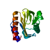
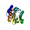
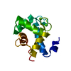
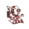
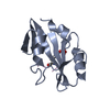
- PDB-1f2k: CRYSTAL STRUCTURE OF ACANTHAMOEBA CASTELLANII PROFILIN II, CUBIC ... -
+ Open data

- Basic information
| Entry | Database: PDB / ID: 1f2k | ||||||
|---|---|---|---|---|---|---|---|
| Title | CRYSTAL STRUCTURE OF ACANTHAMOEBA CASTELLANII PROFILIN II, CUBIC CRYSTAL FORM | ||||||
 Components Components | PROFILIN II | ||||||
 Keywords Keywords | STRUCTURAL PROTEIN / SEVEN-STRANDED INCOMPLETE ANTIPARALLEL UP-AND-DOWN BETA BARREL / ACTIN-BINDING PROTEIN / POLY-L-PROLINE BINDING PROTEIN / PIP2 BINDING PROTEIN | ||||||
| Function / homology |  Function and homology information Function and homology information | ||||||
| Biological species |  Acanthamoeba castellanii (eukaryote) Acanthamoeba castellanii (eukaryote) | ||||||
| Method |  X-RAY DIFFRACTION / SYNCHROTRON / MOLECULAR REPLACEMENT / Resolution: 2.3 Å X-RAY DIFFRACTION / SYNCHROTRON / MOLECULAR REPLACEMENT / Resolution: 2.3 Å | ||||||
 Authors Authors | Fedorov, A.A. / Shi, W. / Mahoney, N. / Kaiser, D.A. / Almo, S.C. | ||||||
 Citation Citation | Journal: To be Published Title: A Comparative Structural Analysis of Profilins Authors: Fedorov, A.A. / Shi, W. / Mahoney, N. / Kaiser, D.A. / Almo, S.C. #1: Journal: Proc.Natl.Acad.Sci.USA / Year: 1994Title: X-ray Structures of Isoforms of the Actin-Binding Protein Profilin that Differ in their Affinity for Phosphatidylinositol Phosphates Authors: Fedorov, A.A. / Magnus, K.A. / Graupe, M.H. / Lattman, E.E. / Pollard, T.D. / Almo, S.C. #2: Journal: J.Mol.Biol. / Year: 1994Title: Purification, Characterization and Crystallization of Human Platelet Profilin Expressed in Escherichia coli Authors: Fedorov, A.A. / Pollard, T.D. / Almo, S.C. #3: Journal: Structure / Year: 1997Title: The Molecular Basis for Allergen Cross-Reactivity: Crystal Structure and IgE-Epitope Mapping of Birch Pollen Profilin Authors: Fedorov, A.A. / Ball, T. / Mahoney, N. / Valenta, R. / Almo, S.C. #4: Journal: J.STRUCT.BIOL. / Year: 1998Title: Crystal Packing Induces a Conformational Change in Profilin-I from Acanthamoeba Castellanii Authors: Liu, S. / Fedorov, A.A. / Pollard, T.D. / Lattman, E.E. / Almo, S.C. / Magnus, K.A. #5: Journal: NAT.STRUCT.BIOL. / Year: 1999Title: Profilin Binds Proline-Rich Ligands in Two Distinct Amido Backbone Orientations Authors: Mahoney, N.M. / Rozwarski, D.A. / Fedorov, E.V. / Fedorov, A.A. / Almo, S.C. | ||||||
| History |
|
- Structure visualization
| Structure viewer | Molecule: MolmilJmol/JSmol |
|---|
- Downloads & links
-Download
| PDBx/mmCIF format | 1f2k.cif.gz | 62.3 KB | Display | PDBx/mmCIF format |
|---|---|---|---|---|
| PDB format | pdb1f2k.ent.gz | 45.9 KB | Display | PDB format |
| PDBx/mmJSON format | 1f2k.json.gz | Tree view | PDBx/mmJSON format | |
| Others |  Other downloads Other downloads |
-Validation report
| Arichive directory | https://data.pdbj.org/pub/pdb/validation_reports/f2/1f2kftp://data.pdbj.org/pub/pdb/validation_reports/f2/1f2k | HTTPS FTP |
|---|
-Related structure data
| Related structure data |  1k0kC  2acgS S: Starting model for refinement C: citing same article ( |
|---|---|
| Similar structure data |


-Links
 PDBj
PDBj
- Assembly
| Deposited unit | 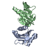
| ||||||||
|---|---|---|---|---|---|---|---|---|---|
| 1 | 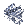
| ||||||||
| 2 | 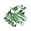
| ||||||||
| Unit cell |
| ||||||||
| Details | The biological assembly is a monomer constructed from chain A or from chain B |
-Components
| #1: Protein | Mass: 12937.442 Da / Num. of mol.: 2 Source method: isolated from a genetically manipulated source Source: (gene. exp.) Acanthamoeba castellanii (eukaryote) / Production host:  #2: Water | ChemComp-HOH / |  Mass: 18.015 Da / Num. of mol.: 179 / Source method: isolated from a natural source / Formula: H2O Mass: 18.015 Da / Num. of mol.: 179 / Source method: isolated from a natural source / Formula: H2O |
|---|
-Experimental details
-Experiment
| Experiment | Method: X-RAY DIFFRACTION / Number of used crystals: 1 |
|---|
- Sample preparation
| Crystal | Density Matthews: 2.64 Å3/Da / Density % sol: 54 % |
|---|---|
| Crystal grow | Temperature: 277 K / Method: vapor diffusion, hanging drop / pH: 5.6 Details: AMMONIUM SULFATE, SODIUM CITRATE, pH 5.6, VAPOR DIFFUSION, HANGING DROP, temperature 277.0K |
-Data collection
| Diffraction | Mean temperature: 140 K |
|---|---|
| Diffraction source | Source: SYNCHROTRON / Site: NSLS  / Beamline: X9B / Wavelength: 1.2 / Beamline: X9B / Wavelength: 1.2 |
| Detector | Type: FUJI / Detector: IMAGE PLATE / Date: Feb 21, 1996 |
| Radiation | Protocol: SINGLE WAVELENGTH / Monochromatic (M) / Laue (L): M / Scattering type: x-ray |
| Radiation wavelength | Wavelength: 1.2 Å / Relative weight: 1 |
| Reflection | Resolution: 2.3→20 Å / Num. all: 13681 / Num. obs: 13681 / % possible obs: 99 % / Observed criterion σ(F): 0 / Observed criterion σ(I): 0 / Redundancy: 4 % / Biso Wilson estimate: 19.2 Å2 / Rmerge(I) obs: 0.073 / Net I/σ(I): 18.3 |
| Reflection shell | Resolution: 2.3→2.4 Å / Redundancy: 3.1 % / Rmerge(I) obs: 0.195 / Mean I/σ(I) obs: 7.2 / % possible all: 99 |
- Processing
| Software |
| ||||||||||||||||||||||||||||||||||||
|---|---|---|---|---|---|---|---|---|---|---|---|---|---|---|---|---|---|---|---|---|---|---|---|---|---|---|---|---|---|---|---|---|---|---|---|---|---|
| Refinement | Method to determine structure: MOLECULAR REPLACEMENT Starting model: 2ACG Resolution: 2.3→20 Å / Isotropic thermal model: RESTRAINED / Cross valid method: THROUGHOUT / σ(F): 2 / Stereochemistry target values: Engh & Huber
| ||||||||||||||||||||||||||||||||||||
| Refine analyze |
| ||||||||||||||||||||||||||||||||||||
| Refinement step | Cycle: LAST / Resolution: 2.3→20 Å
| ||||||||||||||||||||||||||||||||||||
| Refine LS restraints |
| ||||||||||||||||||||||||||||||||||||
| Refine LS restraints NCS | NCS model details: RESTRAINTS | ||||||||||||||||||||||||||||||||||||
| Xplor file | Serial no: 1 / Param file: PROTEIN_REP.PARAM / Topol file: PROTEIN.TOP |
