 Movie
Movie Controller


+ Open data

- Basic information
| Entry | Database: PDB / ID: 1k0k | ||||||
|---|---|---|---|---|---|---|---|
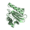
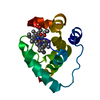
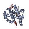
| Title | Yeast Profilin, Cubic Crystal Form | ||||||
 Components Components | PROFILIN | ||||||
 Keywords Keywords | CONTRACTILE PROTEIN / ACTIN-BINDING PROTEIN / PIP2 BINDING PROTEIN / POLY-L-PROLINE BINDING PROTEIN | ||||||
| Function / homology |  Function and homology information Function and homology informationpositive regulation of formin-nucleated actin cable assembly / mitotic actomyosin contractile ring assembly / proline-rich region binding / actin monomer binding / phosphatidylinositol-4,5-bisphosphate binding / protein sequestering activity / cell cortex / cytoskeleton / cytosol Similarity search - Function | ||||||
| Biological species |  | ||||||
| Method |  X-RAY DIFFRACTION / SYNCHROTRON / MOLECULAR REPLACEMENT / Resolution: 2.35 Å X-RAY DIFFRACTION / SYNCHROTRON / MOLECULAR REPLACEMENT / Resolution: 2.35 Å | ||||||
 Authors Authors | Vorobiev, S. / Fedorov, A.A. / Almo, S.C. | ||||||
 Citation Citation | Journal: To be Published Title: A Comparative Structural Analysis of Profilins Authors: Vorobiev, S. / Fedorov, A.A. / Almo, S.C. #1: Journal: Biochemistry / Year: 1998Title: Structure Determination and Characterization of Saccharomyces Cerevisae Profilin Authors: Eads, J.C. / Mahoney, N.M. / Vorobiev, S. / Bresnick, A.R. / Wen, K.K. / Rubenstein, P.A. / Haarer, B.K. / Almo, S.C. #2: Journal: Proc.Natl.Acad.Sci.USA / Year: 1994Title: X-Ray Structures of Isoforms of the Actin-Binding Protein Profilin That Differ in their Affinity for Phosphatidylinositol Phosphates Authors: Fedorov, A.A. / Magnus, K.A. / Graupe, M.H. / Lattman, E.E. / Pollard, T.D. / Almo, S.C. #3: Journal: J.Mol.Biol. / Year: 1994Title: Purification, Characterization and Crystallization of Human Platelet Profilin Expressed in Escherichia Coli Authors: Fedorov, A.A. / Pollard, T.D. / Almo, S.C. #4: Journal: Structure / Year: 1997Title: The Molecular Basis for Allergen Cross-Reactivity: Crystal Structure and Ige-Epitope Mapping of Birch Pollen Profilin Authors: Fedorov, A.A. / Ball, T. / Mahoney, N.M. / Valenta, R. / Almo, S.C. | ||||||
| History |
|
- Structure visualization
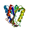
| Structure viewer | Molecule: MolmilJmol/JSmol |
|---|
- Downloads & links
-Download
| PDBx/mmCIF format | 1k0k.cif.gz | 39.4 KB | Display | PDBx/mmCIF format |
|---|---|---|---|---|
| PDB format | pdb1k0k.ent.gz | 27 KB | Display | PDB format |
| PDBx/mmJSON format | 1k0k.json.gz | Tree view | PDBx/mmJSON format | |
| Others |  Other downloads Other downloads |
-Validation report
| Arichive directory | https://data.pdbj.org/pub/pdb/validation_reports/k0/1k0kftp://data.pdbj.org/pub/pdb/validation_reports/k0/1k0k | HTTPS FTP |
|---|
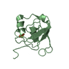
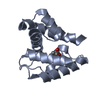
-Related structure data
| Related structure data |  1f2kC 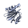 1yprS S: Starting model for refinement C: citing same article ( |
|---|---|
| Similar structure data |


-Links
 PDBj
PDBj
- Assembly
| Deposited unit | 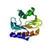
| ||||||||
|---|---|---|---|---|---|---|---|---|---|
| 1 |
| ||||||||
| Unit cell |
| ||||||||
| Details | The biological assembly is a monomer |
-Components
| #1: Protein | Mass: 13559.190 Da / Num. of mol.: 1 Source method: isolated from a genetically manipulated source Source: (gene. exp.) Production host:  |
|---|---|
| #2: Chemical | ChemComp-GOL /   Mass: 92.094 Da / Num. of mol.: 1 / Source method: obtained synthetically / Formula: C3H8O3 Mass: 92.094 Da / Num. of mol.: 1 / Source method: obtained synthetically / Formula: C3H8O3 |
| #3: Water | ChemComp-HOH /  Mass: 18.015 Da / Num. of mol.: 81 / Source method: isolated from a natural source / Formula: H2O Mass: 18.015 Da / Num. of mol.: 81 / Source method: isolated from a natural source / Formula: H2O |
-Experimental details
-Experiment
| Experiment | Method: X-RAY DIFFRACTION / Number of used crystals: 1 |
|---|
- Sample preparation
| Crystal | Density Matthews: 6.165 Å3/Da / Density % sol: 79.28 % |
|---|---|
| Crystal grow | Temperature: 293 K / Method: vapor diffusion, hanging drop / pH: 4.6 Details: sodium format, sodium acetate, pH 4.6, VAPOR DIFFUSION, HANGING DROP, temperature 293.0K |
-Data collection
| Diffraction | Mean temperature: 100 K |
|---|---|
| Diffraction source | Source: SYNCHROTRON / Site: NSLS  / Beamline: X9A / Wavelength: 0.98 Å / Beamline: X9A / Wavelength: 0.98 Å |
| Detector | Type: ADSC QUANTUM 4 / Detector: CCD / Date: Jul 5, 2001 |
| Radiation | Protocol: SINGLE WAVELENGTH / Monochromatic (M) / Laue (L): M / Scattering type: x-ray |
| Radiation wavelength | Wavelength: 0.98 Å / Relative weight: 1 |
| Reflection | Resolution: 2.35→20 Å / Num. all: 14401 / Num. obs: 14401 / % possible obs: 96 % / Observed criterion σ(F): 0 / Observed criterion σ(I): 0 / Biso Wilson estimate: 34.5 Å2 / Rmerge(I) obs: 0.05 / Net I/σ(I): 45.3 |
| Reflection shell | Resolution: 2.35→2.43 Å / Rmerge(I) obs: 0.265 / Mean I/σ(I) obs: 12.9 / % possible all: 92 |
- Processing
| Software |
| ||||||||||||||||||||||||||||||||||||
|---|---|---|---|---|---|---|---|---|---|---|---|---|---|---|---|---|---|---|---|---|---|---|---|---|---|---|---|---|---|---|---|---|---|---|---|---|---|
| Refinement | Method to determine structure: MOLECULAR REPLACEMENT Starting model: PDB ENTRY 1YPR Resolution: 2.35→20 Å / Rfactor Rfree error: 0.006 / Cross valid method: THROUGHOUT / σ(F): 1 / Stereochemistry target values: Engh & Huber
| ||||||||||||||||||||||||||||||||||||
| Solvent computation | Solvent model: flat model / Bsol: 76.5199 Å2 / ksol: 0.391558 e/Å3 | ||||||||||||||||||||||||||||||||||||
| Displacement parameters | Biso mean: 48.2 Å2 | ||||||||||||||||||||||||||||||||||||
| Refine analyze |
| ||||||||||||||||||||||||||||||||||||
| Refinement step | Cycle: LAST / Resolution: 2.35→20 Å
| ||||||||||||||||||||||||||||||||||||
| Refine LS restraints |
| ||||||||||||||||||||||||||||||||||||
| LS refinement shell | Resolution: 2.35→2.5 Å / Rfactor Rfree error: 0.019 / Total num. of bins used: 6
| ||||||||||||||||||||||||||||||||||||
| Xplor file |
|
