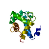
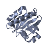
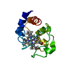
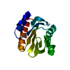
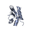
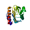
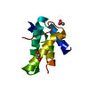
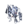
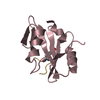
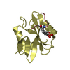
[English] 日本語
Yorodumi
- PDB-5tqs: Phospholipase C gamma-1 C-terminal SH2 domain bound to a phosphop... -

+
Open data


ID or keywords:

Loading...

-
Basic information

Entry
Database: PDB / ID: 5tqs
TitlePhospholipase C gamma-1 C-terminal SH2 domain bound to a phosphopeptide derived from the receptor tyrosine kinase ErbB2
Components
  • 1-phosphatidylinositol 4,5-bisphosphate phosphodiesterase gamma-1
  • Receptor protein-tyrosine kinase
KeywordsHYDROLASE / SH2 / phospholipase / phosphopeptide
Function / homology
Function and homology information


calcium-dependent phospholipase C activity / negative regulation of immature T cell proliferation in thymus / phosphoinositide phospholipase C / ERBB3:ERBB2 complex / ERBB2-ERBB4 signaling pathway / GRB7 events in ERBB2 signaling / immature T cell proliferation in thymus / RNA polymerase I core binding / semaphorin receptor complex / phosphatidylinositol metabolic process ...calcium-dependent phospholipase C activity / negative regulation of immature T cell proliferation in thymus / phosphoinositide phospholipase C / ERBB3:ERBB2 complex / ERBB2-ERBB4 signaling pathway / GRB7 events in ERBB2 signaling / immature T cell proliferation in thymus / RNA polymerase I core binding / semaphorin receptor complex / phosphatidylinositol metabolic process / COP9 signalosome / phosphatidylinositol-4,5-bisphosphate phospholipase C activity / regulation of microtubule-based process / ErbB-3 class receptor binding / motor neuron axon guidance / Sema4D induced cell migration and growth-cone collapse / phospholipid catabolic process / ERBB signaling pathway / neurotransmitter receptor localization to postsynaptic specialization membrane / PLCG1 events in ERBB2 signaling / ERBB2-EGFR signaling pathway / neuromuscular junction development / ERBB2 Activates PTK6 Signaling / enzyme-linked receptor protein signaling pathway / phosphatidylinositol-mediated signaling / positive regulation of Rho protein signal transduction / ERBB2-ERBB3 signaling pathway / Drug-mediated inhibition of ERBB2 signaling / Resistance of ERBB2 KD mutants to trastuzumab / Resistance of ERBB2 KD mutants to sapitinib / Resistance of ERBB2 KD mutants to tesevatinib / Resistance of ERBB2 KD mutants to neratinib / Resistance of ERBB2 KD mutants to osimertinib / Resistance of ERBB2 KD mutants to afatinib / Resistance of ERBB2 KD mutants to AEE788 / Resistance of ERBB2 KD mutants to lapatinib / Drug resistance in ERBB2 TMD/JMD mutants / positive regulation of transcription by RNA polymerase I / oligodendrocyte differentiation / ERBB2 Regulates Cell Motility / semaphorin-plexin signaling pathway / PI3K events in ERBB2 signaling / positive regulation of epithelial cell migration / positive regulation of protein targeting to membrane / cellular response to vascular endothelial growth factor stimulus / regulation of angiogenesis / Schwann cell development / regulation of ERK1 and ERK2 cascade / coreceptor activity / ruffle / release of sequestered calcium ion into cytosol / Signaling by ERBB2 / transmembrane receptor protein tyrosine kinase activity / TFAP2 (AP-2) family regulates transcription of growth factors and their receptors / myelination / positive regulation of MAP kinase activity / GRB2 events in ERBB2 signaling / positive regulation of cell adhesion / SHC1 events in ERBB2 signaling / cellular response to epidermal growth factor stimulus / Downregulation of ERBB2:ERBB3 signaling / Constitutive Signaling by Overexpressed ERBB2 / cell surface receptor protein tyrosine kinase signaling pathway / guanyl-nucleotide exchange factor activity / positive regulation of epithelial cell proliferation / basal plasma membrane / positive regulation of translation / phosphatidylinositol 3-kinase/protein kinase B signal transduction / wound healing / Signaling by ERBB2 TMD/JMD mutants / placental growth factor receptor activity / insulin receptor activity / vascular endothelial growth factor receptor activity / hepatocyte growth factor receptor activity / macrophage colony-stimulating factor receptor activity / platelet-derived growth factor alpha-receptor activity / platelet-derived growth factor beta-receptor activity / stem cell factor receptor activity / boss receptor activity / protein tyrosine kinase collagen receptor activity / brain-derived neurotrophic factor receptor activity / transmembrane-ephrin receptor activity / GPI-linked ephrin receptor activity / epidermal growth factor receptor activity / fibroblast growth factor receptor activity / insulin-like growth factor receptor activity / Signaling by ERBB2 ECD mutants / receptor protein-tyrosine kinase / neuromuscular junction / cellular response to growth factor stimulus / peptidyl-tyrosine phosphorylation / Signaling by ERBB2 KD Mutants / receptor tyrosine kinase binding / epidermal growth factor receptor signaling pathway / ruffle membrane / Downregulation of ERBB2 signaling / neuron differentiation / Constitutive Signaling by Aberrant PI3K in Cancer / transmembrane signaling receptor activity / PIP3 activates AKT signaling
Similarity search - Function
1-phosphatidylinositol 4,5-bisphosphate phosphodiesterase gamma-1, SH3 domain / PLCG EF-hand motif 1 / Phosphatidylinositol-4, 5-bisphosphate phosphodiesterase gamma / PLC-gamma, C-terminal SH2 domain / PLC-gamma, N-terminal SH2 domain / PLCG EF-hand motif 2 / Phosphoinositide phospholipase C family / Phospholipase C, phosphatidylinositol-specific, Y domain / Phosphatidylinositol-specific phospholipase C, Y domain / Phosphatidylinositol-specific phospholipase Y-box domain profile. ...1-phosphatidylinositol 4,5-bisphosphate phosphodiesterase gamma-1, SH3 domain / PLCG EF-hand motif 1 / Phosphatidylinositol-4, 5-bisphosphate phosphodiesterase gamma / PLC-gamma, C-terminal SH2 domain / PLC-gamma, N-terminal SH2 domain / PLCG EF-hand motif 2 / Phosphoinositide phospholipase C family / Phospholipase C, phosphatidylinositol-specific, Y domain / Phosphatidylinositol-specific phospholipase C, Y domain / Phosphatidylinositol-specific phospholipase Y-box domain profile. / Phospholipase C, catalytic domain (part); domain Y / Phosphatidylinositol-specific phospholipase C, X domain / Phosphatidylinositol-specific phospholipase C, X domain / Phospholipase C, catalytic domain (part); domain X / Phosphatidylinositol-specific phospholipase X-box domain profile. / PLC-like phosphodiesterase, TIM beta/alpha-barrel domain superfamily / : / Epidermal growth factor receptor transmembrane-juxtamembrane segment / SH2 domain / Tyrosine protein kinase, EGF/ERB/XmrK receptor / Growth factor receptor domain 4 / Growth factor receptor domain IV / SHC Adaptor Protein / Protein kinase C conserved region 2 (CalB) / C2 domain / C2 domain / C2 domain profile. / Receptor L-domain / Furin-like cysteine-rich domain / Receptor L-domain superfamily / Furin-like cysteine rich region / Receptor L domain / Furin-like repeat / Furin-like repeats / PH domain / C2 domain superfamily / PH domain profile. / Pleckstrin homology domain. / Growth factor receptor cysteine-rich domain superfamily / Pleckstrin homology domain / : / SH3 domain / SH2 domain / Src homology 2 (SH2) domain profile. / Src homology 2 domains / SH2 domain / SH2 domain superfamily / Src homology 3 domains / EF-Hand 1, calcium-binding site / SH3-like domain superfamily / EF-hand calcium-binding domain. / Src homology 3 (SH3) domain profile. / SH3 domain / EF-hand calcium-binding domain profile. / EF-hand domain / EF-hand domain pair / Tyrosine-protein kinase, catalytic domain / Tyrosine kinase, catalytic domain / Tyrosine protein kinases specific active-site signature. / Tyrosine-protein kinase, active site / PH-like domain superfamily / Serine-threonine/tyrosine-protein kinase, catalytic domain / Protein tyrosine and serine/threonine kinase / Protein kinase, ATP binding site / Protein kinases ATP-binding region signature. / Protein kinase domain profile. / Protein kinase domain / Protein kinase-like domain superfamily / 2-Layer Sandwich / Alpha Beta
Similarity search - Domain/homology
Receptor tyrosine-protein kinase erbB-2 / Receptor tyrosine-protein kinase erbB-2 / 1-phosphatidylinositol 4,5-bisphosphate phosphodiesterase gamma-1
Similarity search - Component
Biological speciesBos taurus (domestic cattle)
Homo sapiens (human)
MethodX-RAY DIFFRACTION / SYNCHROTRON / MOLECULAR REPLACEMENT / molecular replacement / Resolution: 1.876 Å
AuthorsWuttke, D.S. / McKercher, M.A.
Funding support United States, 1items
OrganizationGrant numberCountry
National Science Foundation (NSF, United States)MCB1121842 United States
CitationJournal: Biochemistry / Year: 2017
Title: Multimodal Recognition of Diverse Peptides by the C-Terminal SH2 Domain of Phospholipase C-gamma 1 Protein.
Authors: McKercher, M.A. / Guan, X. / Tan, Z. / Wuttke, D.S.
History
DepositionOct 24, 2016Deposition site: RCSB / Processing site: RCSB
Revision 1.0Apr 19, 2017Provider: repository / Type: Initial release
Revision 1.1Apr 26, 2017Group: Database references
Revision 1.2May 10, 2017Group: Database references
Revision 1.3Sep 27, 2017Group: Author supporting evidence / Category: pdbx_audit_support / Item: _pdbx_audit_support.funding_organization
Revision 1.4Nov 27, 2019Group: Author supporting evidence / Category: pdbx_audit_support / Item: _pdbx_audit_support.funding_organization
Revision 1.5Oct 4, 2023Group: Data collection / Database references / Refinement description
Category: chem_comp_atom / chem_comp_bond ...chem_comp_atom / chem_comp_bond / database_2 / pdbx_initial_refinement_model
Item: _database_2.pdbx_DOI / _database_2.pdbx_database_accession
Revision 1.6Nov 15, 2023Group: Data collection / Category: chem_comp_atom / chem_comp_bond / Item: _chem_comp_atom.atom_id / _chem_comp_bond.atom_id_2
Revision 1.7Nov 6, 2024Group: Structure summary / Category: pdbx_entry_details / pdbx_modification_feature

-
Structure visualization

Structure viewerMolecule:
MolmilJmol/JSmol

Downloads & links

-
Assembly

Deposited unit
A: 1-phosphatidylinositol 4,5-bisphosphate phosphodiesterase gamma-1
B: 1-phosphatidylinositol 4,5-bisphosphate phosphodiesterase gamma-1
C: 1-phosphatidylinositol 4,5-bisphosphate phosphodiesterase gamma-1
D: 1-phosphatidylinositol 4,5-bisphosphate phosphodiesterase gamma-1
E: Receptor protein-tyrosine kinase
F: Receptor protein-tyrosine kinase
H: Receptor protein-tyrosine kinase
G: Receptor protein-tyrosine kinase


Theoretical massNumber of molelcules
Total (without water)53,9808
Polymers53,9808
Non-polymers00
Water4,666259
1
A: 1-phosphatidylinositol 4,5-bisphosphate phosphodiesterase gamma-1
E: Receptor protein-tyrosine kinase


Theoretical massNumber of molelcules
Total (without water)13,4952
Polymers13,4952
Non-polymers00
Water362
TypeNameSymmetry operationNumber
identity operation1_555x,y,z1
Buried area790 Å2
ΔGint-7 kcal/mol
Surface area6200 Å2
MethodPISA
2
B: 1-phosphatidylinositol 4,5-bisphosphate phosphodiesterase gamma-1
F: Receptor protein-tyrosine kinase


Theoretical massNumber of molelcules
Total (without water)13,4952
Polymers13,4952
Non-polymers00
Water362
TypeNameSymmetry operationNumber
identity operation1_555x,y,z1
Buried area800 Å2
ΔGint-7 kcal/mol
Surface area6380 Å2
MethodPISA
3
C: 1-phosphatidylinositol 4,5-bisphosphate phosphodiesterase gamma-1
G: Receptor protein-tyrosine kinase


Theoretical massNumber of molelcules
Total (without water)13,4952
Polymers13,4952
Non-polymers00
Water362
TypeNameSymmetry operationNumber
identity operation1_555x,y,z1
Buried area730 Å2
ΔGint-6 kcal/mol
Surface area6380 Å2
MethodPISA
4
D: 1-phosphatidylinositol 4,5-bisphosphate phosphodiesterase gamma-1
H: Receptor protein-tyrosine kinase


Theoretical massNumber of molelcules
Total (without water)13,4952
Polymers13,4952
Non-polymers00
Water362
TypeNameSymmetry operationNumber
identity operation1_555x,y,z1
Buried area790 Å2
ΔGint-6 kcal/mol
Surface area6580 Å2
MethodPISA
Unit cell
Length a, b, c (Å)65.320, 30.620, 104.570
Angle α, β, γ (deg.)90.000, 100.120, 90.000
Int Tables number4
Space group name H-MP1211

-
Components

#1: Protein
1-phosphatidylinositol 4,5-bisphosphate phosphodiesterase gamma-1 / PLC-148 / Phosphoinositide phospholipase C-gamma-1 / Phospholipase C-II / PLC-II / Phospholipase C- ...PLC-148 / Phosphoinositide phospholipase C-gamma-1 / Phospholipase C-II / PLC-II / Phospholipase C-gamma-1 / PLC-gamma-1


Mass: 11989.685 Da / Num. of mol.: 4 / Fragment: UNP residues 663-759
Source method: isolated from a genetically manipulated source
Source: (gene. exp.) Bos taurus (domestic cattle) / Gene: PLCG1 / Plasmid: pET15b / Cell line (production host): BL21(DE3) / Production host: Escherichia coli (E. coli)
References: UniProt: P08487, phosphoinositide phospholipase C
#2: Protein/peptide
Receptor protein-tyrosine kinase


Mass: 1505.434 Da / Num. of mol.: 4 / Fragment: UNP residues 942-952 / Source method: obtained synthetically / Source: (synth.) Homo sapiens (human) / References: UniProt: B4DTR1, UniProt: P04626*PLUS
#3: Water ChemComp-HOH / water


Mass: 18.015 Da / Num. of mol.: 259 / Source method: isolated from a natural source / Formula: H2O
Has protein modificationY

-
Experimental details

-
Experiment

ExperimentMethod: X-RAY DIFFRACTION / Number of used crystals: 1

-
Sample preparation

CrystalDensity Matthews: 1.95 Å3/Da / Density % sol: 36.98 %
Crystal growTemperature: 277 K / Method: vapor diffusion, hanging drop / Details: 0.1 M sodium acetate (pH 5.5), 22% PEG MME 5000

-
Data collection

DiffractionMean temperature: 100 K
Diffraction sourceSource: SYNCHROTRON / Site: ALS / Beamline: 8.2.1 / Wavelength: 1 Å
DetectorType: ADSC QUANTUM 315r / Detector: CCD / Date: Aug 4, 2016
RadiationProtocol: SINGLE WAVELENGTH / Monochromatic (M) / Laue (L): M / Scattering type: x-ray
Radiation wavelengthWavelength: 1 Å / Relative weight: 1
ReflectionResolution: 1.876→64.304 Å / Num. obs: 33698 / % possible obs: 99 % / Redundancy: 3.4 % / Biso Wilson estimate: 25.07 Å2 / Rpim(I) all: 0.055 / Rrim(I) all: 0.102 / Rsym value: 0.086 / Net I/av σ(I): 4.902 / Net I/σ(I): 9.1 / Num. measured all: 112903
Reflection shell
Resolution (Å)Redundancy (%)Rmerge(I) obsMean I/σ(I) obsDiffraction-ID% possible all
1.876-1.982.80.4331.5196.3
1.98-2.13.10.2892.3199.1
2.1-2.243.60.1923.5199.4
2.24-2.423.60.1394.9199.5
2.42-2.653.60.1026.4199.6
2.65-2.973.60.0847.6199.6
2.97-3.433.50.0698.5199.7
3.43-4.23.40.0688199.8
4.2-5.933.30.0697.8199.5
5.93-64.3043.20.0578.6199

-
Phasing

PhasingMethod: molecular replacement
Phasing MRModel details: Phaser MODE: MR_AUTO
Highest resolutionLowest resolution
Rotation1.88 Å64.3 Å
Translation1.88 Å64.3 Å

-
Processing

Software
NameVersionClassification
SCALA3.3.22data scaling
PHASER2.6.1phasing
PHENIX1.10.1_2155refinement
PDB_EXTRACT3.2data extraction
MOSFLMdata reduction
RefinementMethod to determine structure: MOLECULAR REPLACEMENT
Starting model: 4K44
Resolution: 1.876→51.472 Å / SU ML: 0.23 / Cross valid method: THROUGHOUT / σ(F): 1.35 / Phase error: 23.69
RfactorNum. reflection% reflection
Rfree0.237 1684 5 %
Rwork0.1885 --
obs0.1909 33684 98.62 %
Solvent computationShrinkage radii: 0.9 Å / VDW probe radii: 1.11 Å
Displacement parametersBiso max: 153.7 Å2 / Biso mean: 36.9937 Å2 / Biso min: 9.6 Å2
Refinement stepCycle: final / Resolution: 1.876→51.472 Å
ProteinNucleic acidLigandSolventTotal
Num. atoms3531 0 0 268 3799
Biso mean---36.12 -
Num. residues----426
Refine LS restraints
Refine-IDTypeDev idealNumber
X-RAY DIFFRACTIONf_bond_d0.0073623
X-RAY DIFFRACTIONf_angle_d0.824875
X-RAY DIFFRACTIONf_chiral_restr0.049479
X-RAY DIFFRACTIONf_plane_restr0.005625
X-RAY DIFFRACTIONf_dihedral_angle_d15.5332171
LS refinement shell

Refine-ID: X-RAY DIFFRACTION / Total num. of bins used: 12

Resolution (Å)Rfactor RfreeNum. reflection RfreeRfactor RworkNum. reflection RworkNum. reflection all% reflection obs (%)
1.8762-1.93140.29991300.26062505263594
1.9314-1.99380.31491390.23252618275798
1.9938-2.0650.28581390.21692637277699
2.065-2.14770.2511380.2042628276699
2.1477-2.24550.23081410.19732677281899
2.2455-2.36380.2551400.20222663280399
2.3638-2.51190.28951400.1962653279399
2.5119-2.70590.22631390.19432658279799
2.7059-2.97820.24421430.19672703284699
2.9782-3.4090.20051430.176327192862100
3.409-4.29470.22661430.156527162859100
4.2947-51.49090.22251490.18712823297299

+
About Yorodumi

-
News

-
Feb 9, 2022. New format data for meta-information of EMDB entries

New format data for meta-information of EMDB entries

  • Version 3 of the EMDB header file is now the official format.
  • The previous official version 1.9 will be removed from the archive.

Related info.:EMDB header

External links:wwPDB to switch to version 3 of the EMDB data model

-
Aug 12, 2020. Covid-19 info

Covid-19 info

URL: https://pdbj.org/emnavi/covid19.php

New page: Covid-19 featured information page in EM Navigator.

Related info.:Covid-19 info / Mar 5, 2020. Novel coronavirus structure data

+
Mar 5, 2020. Novel coronavirus structure data

Novel coronavirus structure data

Related info.:Yorodumi Speices / Aug 12, 2020. Covid-19 info

External links:COVID-19 featured content - PDBj / Molecule of the Month (242):Coronavirus Proteases

+
Jan 31, 2019. EMDB accession codes are about to change! (news from PDBe EMDB page)

EMDB accession codes are about to change! (news from PDBe EMDB page)

  • The allocation of 4 digits for EMDB accession codes will soon come to an end. Whilst these codes will remain in use, new EMDB accession codes will include an additional digit and will expand incrementally as the available range of codes is exhausted. The current 4-digit format prefixed with “EMD-” (i.e. EMD-XXXX) will advance to a 5-digit format (i.e. EMD-XXXXX), and so on. It is currently estimated that the 4-digit codes will be depleted around Spring 2019, at which point the 5-digit format will come into force.
  • The EM Navigator/Yorodumi systems omit the EMD- prefix.

Related info.:Q: What is EMD? / ID/Accession-code notation in Yorodumi/EM Navigator

External links:EMDB Accession Codes are Changing Soon! / Contact to PDBj

+
Jul 12, 2017. Major update of PDB

Major update of PDB

  • wwPDB released updated PDB data conforming to the new PDBx/mmCIF dictionary.
  • This is a major update changing the version number from 4 to 5, and with Remediation, in which all the entries are updated.
  • In this update, many items about electron microscopy experimental information are reorganized (e.g. em_software).
  • Now, EM Navigator and Yorodumi are based on the updated data.

External links:wwPDB Remediation / Enriched Model Files Conforming to OneDep Data Standards Now Available in the PDB FTP Archive

-
Yorodumi

Thousand views of thousand structures

  • Yorodumi is a browser for structure data from EMDB, PDB, SASBDB, etc.
  • This page is also the successor to EM Navigator detail page, and also detail information page/front-end page for Omokage search.
  • The word "yorodu" (or yorozu) is an old Japanese word meaning "ten thousand". "mi" (miru) is to see.

Related info.:EMDB / PDB / SASBDB / Comparison of 3 databanks / Yorodumi Search / Aug 31, 2016. New EM Navigator & Yorodumi / Yorodumi Papers / Jmol/JSmol / Function and homology information / Changes in new EM Navigator and Yorodumi

Read more