 Movie
Movie Controller

[English] 日本語
 Yorodumi
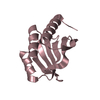
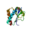
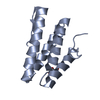
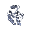
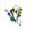
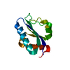
- PDB-5to4: Phospholipase C gamma-1 C-terminal SH2 domain, spacegroup P212121 -
+ Open data

- Basic information
| Entry | Database: PDB / ID: 5to4 | ||||||
|---|---|---|---|---|---|---|---|
| Title | Phospholipase C gamma-1 C-terminal SH2 domain, spacegroup P212121 | ||||||
 Components Components | 1-phosphatidylinositol 4,5-bisphosphate phosphodiesterase gamma-1 | ||||||
 Keywords Keywords | HYDROLASE / SH2 / Phospholipase | ||||||
| Function / homology |  Function and homology information Function and homology information: / phosphatidylinositol phospholipase C activity / phosphoinositide phospholipase C / phosphatidylinositol metabolic process / phosphatidylinositol-4,5-bisphosphate phospholipase C activity / COP9 signalosome / phospholipid catabolic process / phosphatidylinositol-mediated signaling / positive regulation of epithelial cell migration / cellular response to vascular endothelial growth factor stimulus ...: / phosphatidylinositol phospholipase C activity / phosphoinositide phospholipase C / phosphatidylinositol metabolic process / phosphatidylinositol-4,5-bisphosphate phospholipase C activity / COP9 signalosome / phospholipid catabolic process / phosphatidylinositol-mediated signaling / positive regulation of epithelial cell migration / cellular response to vascular endothelial growth factor stimulus / release of sequestered calcium ion into cytosol / ruffle / guanyl-nucleotide exchange factor activity / cellular response to epidermal growth factor stimulus / epidermal growth factor receptor signaling pathway / ruffle membrane / lamellipodium / in utero embryonic development / calcium ion binding / plasma membrane / cytoplasm Similarity search - Function | ||||||
| Biological species |  | ||||||
| Method |  X-RAY DIFFRACTION / SYNCHROTRON / MOLECULAR REPLACEMENT / molecular replacement / Resolution: 1.7 Å X-RAY DIFFRACTION / SYNCHROTRON / MOLECULAR REPLACEMENT / molecular replacement / Resolution: 1.7 Å | ||||||
 Authors Authors | Wuttke, D.S. / McKercher, M.A. | ||||||
 Citation Citation | Journal: Biochemistry / Year: 2017 Title: Multimodal Recognition of Diverse Peptides by the C-Terminal SH2 Domain of Phospholipase C-gamma 1 Protein. Authors: McKercher, M.A. / Guan, X. / Tan, Z. / Wuttke, D.S. | ||||||
| History |
|
- Structure visualization
| Structure viewer | Molecule: MolmilJmol/JSmol |
|---|
- Downloads & links
-Download
| PDBx/mmCIF format | 5to4.cif.gz | 56.1 KB | Display | PDBx/mmCIF format |
|---|---|---|---|---|
| PDB format | pdb5to4.ent.gz | 39.4 KB | Display | PDB format |
| PDBx/mmJSON format | 5to4.json.gz | Tree view | PDBx/mmJSON format | |
| Others |  Other downloads Other downloads |
-Validation report
| Arichive directory | https://data.pdbj.org/pub/pdb/validation_reports/to/5to4ftp://data.pdbj.org/pub/pdb/validation_reports/to/5to4 | HTTPS FTP |
|---|
-Related structure data
| Related structure data |  5tnwC  5tq1C  5tqsC  4k44S C: citing same article ( S: Starting model for refinement |
|---|---|
| Similar structure data |


-Links
 PDBj
PDBj


- Assembly
| Deposited unit | 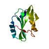
| ||||||||
|---|---|---|---|---|---|---|---|---|---|
| 1 |
| ||||||||
| Unit cell |
|
-Components
| #1: Protein | Mass: 11989.685 Da / Num. of mol.: 1 / Fragment: UNP residues 663-759 Source method: isolated from a genetically manipulated source Details: Scattered electron density was also present for at least two additional polymers, likely low molecular weight polyethylene glycol molecules. However, the molecules could not be fit into the ...Details: Scattered electron density was also present for at least two additional polymers, likely low molecular weight polyethylene glycol molecules. However, the molecules could not be fit into the density non-ambiguously and were consequently omitted from the model. Source: (gene. exp.)  References: UniProt: P08487, phosphoinositide phospholipase C |
|---|---|
| #2: Water | ChemComp-HOH /  Mass: 18.015 Da / Num. of mol.: 74 / Source method: isolated from a natural source / Formula: H2O Mass: 18.015 Da / Num. of mol.: 74 / Source method: isolated from a natural source / Formula: H2O |
-Experimental details
-Experiment
| Experiment | Method: X-RAY DIFFRACTION / Number of used crystals: 1 |
|---|
- Sample preparation
| Crystal | Density Matthews: 2.32 Å3/Da / Density % sol: 47.01 % |
|---|---|
| Crystal grow | Temperature: 277 K / Method: vapor diffusion, hanging drop / Details: 0.1 M sodium citrate (pH 5.5), 22% PEG 1000 |
-Data collection
| Diffraction | Mean temperature: 100 K | ||||||||||||||||||||||||||||||||||||||||||||||||||||||||||||||||||
|---|---|---|---|---|---|---|---|---|---|---|---|---|---|---|---|---|---|---|---|---|---|---|---|---|---|---|---|---|---|---|---|---|---|---|---|---|---|---|---|---|---|---|---|---|---|---|---|---|---|---|---|---|---|---|---|---|---|---|---|---|---|---|---|---|---|---|---|
| Diffraction source | Source: SYNCHROTRON / Site: ALS  / Beamline: 8.2.1 / Wavelength: 1 Å / Beamline: 8.2.1 / Wavelength: 1 Å | ||||||||||||||||||||||||||||||||||||||||||||||||||||||||||||||||||
| Detector | Type: ADSC QUANTUM 315r / Detector: CCD / Date: May 4, 2016 | ||||||||||||||||||||||||||||||||||||||||||||||||||||||||||||||||||
| Radiation | Protocol: SINGLE WAVELENGTH / Monochromatic (M) / Laue (L): M / Scattering type: x-ray | ||||||||||||||||||||||||||||||||||||||||||||||||||||||||||||||||||
| Radiation wavelength | Wavelength: 1 Å / Relative weight: 1 | ||||||||||||||||||||||||||||||||||||||||||||||||||||||||||||||||||
| Reflection | Resolution: 1.7→36.44 Å / Num. all: 12342 / Num. obs: 12342 / % possible obs: 97.9 % / Redundancy: 6.5 % / Biso Wilson estimate: 20.08 Å2 / Rpim(I) all: 0.036 / Rrim(I) all: 0.092 / Rsym value: 0.084 / Net I/av σ(I): 4.706 / Net I/σ(I): 12.8 / Num. measured all: 80381 | ||||||||||||||||||||||||||||||||||||||||||||||||||||||||||||||||||
| Reflection shell |
|
-Phasing
| Phasing | Method: molecular replacement | |||||||||
|---|---|---|---|---|---|---|---|---|---|---|
| Phasing MR | Model details: Phaser MODE: MR_AUTO
|
- Processing
| Software |
| |||||||||||||||||||||||||||||||||||
|---|---|---|---|---|---|---|---|---|---|---|---|---|---|---|---|---|---|---|---|---|---|---|---|---|---|---|---|---|---|---|---|---|---|---|---|---|
| Refinement | Method to determine structure: MOLECULAR REPLACEMENT Starting model: 4K44 Resolution: 1.7→28.114 Å / SU ML: 0.18 / Cross valid method: THROUGHOUT / σ(F): 1.34 / Phase error: 23.84
| |||||||||||||||||||||||||||||||||||
| Solvent computation | Shrinkage radii: 0.9 Å / VDW probe radii: 1.11 Å | |||||||||||||||||||||||||||||||||||
| Displacement parameters | Biso max: 100.19 Å2 / Biso mean: 29.6369 Å2 / Biso min: 10.26 Å2 | |||||||||||||||||||||||||||||||||||
| Refinement step | Cycle: final / Resolution: 1.7→28.114 Å
| |||||||||||||||||||||||||||||||||||
| Refine LS restraints |
| |||||||||||||||||||||||||||||||||||
| LS refinement shell | Refine-ID: X-RAY DIFFRACTION / Total num. of bins used: 4
|
