 Movie
Movie Controller

[English] 日本語
 Yorodumi
- PDB-3jxt: Crystal structure of the third PDZ domain of SAP-102 in complex w... -
+ Open data

- Basic information
| Entry | Database: PDB / ID: 3jxt | ||||||
|---|---|---|---|---|---|---|---|
| Title | Crystal structure of the third PDZ domain of SAP-102 in complex with a fluorogenic peptide-based ligand | ||||||
 Components Components |
| ||||||
 Keywords Keywords | SIGNALING PROTEIN / SAP102 / DLG3 / Stargazin / 4-DMAP / 4DB / PDZ domain / solvatochromic flurophore / fluorogenic probe / Calcium channel / Calcium transport / Ion transport / Ionic channel / Transport / Voltage-gated channel | ||||||
| Function / homology |  Function and homology information Function and homology informationestablishment of planar polarity / Presynaptic depolarization and calcium channel opening / Neurexins and neuroligins / LGI-ADAM interactions / structural constituent of postsynaptic density / Trafficking of AMPA receptors / receptor localization to synapse / eye blink reflex / positive regulation of protein localization to basolateral plasma membrane / establishment or maintenance of epithelial cell apical/basal polarity ...establishment of planar polarity / Presynaptic depolarization and calcium channel opening / Neurexins and neuroligins / LGI-ADAM interactions / structural constituent of postsynaptic density / Trafficking of AMPA receptors / receptor localization to synapse / eye blink reflex / positive regulation of protein localization to basolateral plasma membrane / establishment or maintenance of epithelial cell apical/basal polarity / cerebellar mossy fiber / protein localization to synapse / postsynaptic neurotransmitter receptor diffusion trapping / regulation of AMPA receptor activity / Activation of Ca-permeable Kainate Receptor / membrane hyperpolarization / channel regulator activity / RAF/MAP kinase cascade / nervous system process / Synaptic adhesion-like molecules / protein targeting to membrane / voltage-gated calcium channel complex / neurotransmitter receptor localization to postsynaptic specialization membrane / receptor clustering / transmission of nerve impulse / neuromuscular junction development / AMPA glutamate receptor complex / membrane depolarization / bicellular tight junction / Unblocking of NMDA receptors, glutamate binding and activation / regulation of postsynaptic membrane neurotransmitter receptor levels / phosphatase binding / voltage-gated calcium channel activity / postsynaptic density, intracellular component / positive regulation of synaptic transmission, glutamatergic / somatodendritic compartment / ionotropic glutamate receptor binding / hippocampal mossy fiber to CA3 synapse / dendritic shaft / PDZ domain binding / neuromuscular junction / regulation of membrane potential / cell-cell adhesion / response to calcium ion / postsynaptic density membrane / Schaffer collateral - CA1 synapse / kinase binding / cell-cell junction / nervous system development / growth cone / protein phosphatase binding / chemical synaptic transmission / basolateral plasma membrane / neuron projection / postsynaptic density / protein domain specific binding / neuronal cell body / ubiquitin protein ligase binding / synapse / protein kinase binding / glutamatergic synapse / cell surface / plasma membrane / cytoplasm Similarity search - Function | ||||||
| Biological species |  | ||||||
| Method |  X-RAY DIFFRACTION / SYNCHROTRON / MOLECULAR REPLACEMENT / Resolution: 1.5 Å X-RAY DIFFRACTION / SYNCHROTRON / MOLECULAR REPLACEMENT / Resolution: 1.5 Å | ||||||
 Authors Authors | Sainlos, M. / Olivier, N.B. / Imperiali, B. | ||||||
 Citation Citation | Journal: J.Am.Chem.Soc. / Year: 2013 Title: Caged mono- and divalent ligands for light-assisted disruption of PDZ domain-mediated interactions. Authors: Sainlos, M. / Iskenderian-Epps, W.S. / Olivier, N.B. / Choquet, D. / Imperiali, B. | ||||||
| History |
|
- Structure visualization
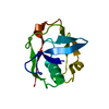
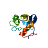
| Structure viewer | Molecule: MolmilJmol/JSmol |
|---|
- Downloads & links
-Download
| PDBx/mmCIF format | 3jxt.cif.gz | 60.7 KB | Display | PDBx/mmCIF format |
|---|---|---|---|---|
| PDB format | pdb3jxt.ent.gz | 43.7 KB | Display | PDB format |
| PDBx/mmJSON format | 3jxt.json.gz | Tree view | PDBx/mmJSON format | |
| Others |  Other downloads Other downloads |
-Validation report
| Arichive directory | https://data.pdbj.org/pub/pdb/validation_reports/jx/3jxtftp://data.pdbj.org/pub/pdb/validation_reports/jx/3jxt | HTTPS FTP |
|---|
-Related structure data
| Related structure data |  3jvq S: Starting model for refinement |
|---|---|
| Similar structure data |


-Links
 PDBj
PDBj


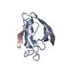
- Assembly
| Deposited unit | 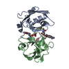
| ||||||||
|---|---|---|---|---|---|---|---|---|---|
| 1 | 
| ||||||||
| 2 | 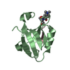
| ||||||||
| Unit cell |
| ||||||||
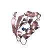
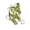
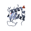
| Details | The biological assembly is the monomeric complex composed of one PDZ domain and its ligand. |
-Components
| #1: Protein | Mass: 11145.364 Da / Num. of mol.: 2 / Fragment: Third PDZ domain: UNP residues 393-493 Source method: isolated from a genetically manipulated source Source: (gene. exp.)  #2: Protein/peptide | Mass: 872.987 Da / Num. of mol.: 2 Fragment: C-terminal motif of Stargazin: UNP O88602 residues 318-323 Mutation: R318(4DB) / Source method: obtained synthetically Details: Peptide ligand obtained by solid phase peptide synthesis References: UniProt: O88602 #3: Chemical | ChemComp-ACT / |   Mass: 59.044 Da / Num. of mol.: 1 / Source method: obtained synthetically / Formula: C2H3O2 Mass: 59.044 Da / Num. of mol.: 1 / Source method: obtained synthetically / Formula: C2H3O2#4: Water | ChemComp-HOH / |  Mass: 18.015 Da / Num. of mol.: 340 / Source method: isolated from a natural source / Formula: H2O Mass: 18.015 Da / Num. of mol.: 340 / Source method: isolated from a natural source / Formula: H2OHas protein modification | Y | |
|---|
-Experimental details
-Experiment
| Experiment | Method: X-RAY DIFFRACTION / Number of used crystals: 1 |
|---|
- Sample preparation
| Crystal | Density Matthews: 2.41 Å3/Da / Density % sol: 48.87 % |
|---|---|
| Crystal grow | Temperature: 277 K / Method: vapor diffusion, hanging drop / pH: 8.5 Details: 1.0 M Sodium citrate, 0.1 M Tris-HCl pH 8.5, VAPOR DIFFUSION, HANGING DROP, temperature 277K |
-Data collection
| Diffraction | Mean temperature: 100 K |
|---|---|
| Diffraction source | Source: SYNCHROTRON / Site: NSLS  / Beamline: X6A / Wavelength: 0.97 Å / Beamline: X6A / Wavelength: 0.97 Å |
| Detector | Type: ADSC QUANTUM 270 / Detector: CCD / Date: Feb 7, 2009 / Details: Toroidal focusing mirror |
| Radiation | Monochromator: Si(111) channel cut / Protocol: SINGLE WAVELENGTH / Monochromatic (M) / Laue (L): M / Scattering type: x-ray |
| Radiation wavelength | Wavelength: 0.97 Å / Relative weight: 1 |
| Reflection | Resolution: 1.5→50 Å / Num. all: 37896 / Num. obs: 37820 / % possible obs: 99.8 % / Observed criterion σ(F): 0 / Observed criterion σ(I): -3 / Redundancy: 7.2 % / Biso Wilson estimate: 16.346 Å2 / Rmerge(I) obs: 0.061 / Net I/σ(I): 28.19 |
| Reflection shell | Resolution: 1.5→1.55 Å / Redundancy: 7.1 % / Rmerge(I) obs: 0.24 / Mean I/σ(I) obs: 6.94 / Num. unique all: 3720 / Rsym value: 0.24 / % possible all: 100 |
- Processing
| Software |
| ||||||||||||||||||||||||||||||||||||||||||||||||||||||||||||||||||||||||||||||||||||||||||||||||||||||||||||||||||||||||||||||||||||||||||||||||||||||||||||||||||||||||||
|---|---|---|---|---|---|---|---|---|---|---|---|---|---|---|---|---|---|---|---|---|---|---|---|---|---|---|---|---|---|---|---|---|---|---|---|---|---|---|---|---|---|---|---|---|---|---|---|---|---|---|---|---|---|---|---|---|---|---|---|---|---|---|---|---|---|---|---|---|---|---|---|---|---|---|---|---|---|---|---|---|---|---|---|---|---|---|---|---|---|---|---|---|---|---|---|---|---|---|---|---|---|---|---|---|---|---|---|---|---|---|---|---|---|---|---|---|---|---|---|---|---|---|---|---|---|---|---|---|---|---|---|---|---|---|---|---|---|---|---|---|---|---|---|---|---|---|---|---|---|---|---|---|---|---|---|---|---|---|---|---|---|---|---|---|---|---|---|---|---|---|---|
| Refinement | Method to determine structure: MOLECULAR REPLACEMENT Starting model: PDB entry 3JVQ 3jvq Resolution: 1.5→45.87 Å / Cor.coef. Fo:Fc: 0.96 / Cor.coef. Fo:Fc free: 0.946 / Cross valid method: THROUGHOUT / ESU R: 0.073 / ESU R Free: 0.075 / Stereochemistry target values: MAXIMUM LIKELIHOOD / Details: HYDROGENS HAVE BEEN ADDED IN THE RIDING POSITIONS
| ||||||||||||||||||||||||||||||||||||||||||||||||||||||||||||||||||||||||||||||||||||||||||||||||||||||||||||||||||||||||||||||||||||||||||||||||||||||||||||||||||||||||||
| Solvent computation | Ion probe radii: 0.8 Å / Shrinkage radii: 0.8 Å / VDW probe radii: 1.2 Å / Solvent model: MASK | ||||||||||||||||||||||||||||||||||||||||||||||||||||||||||||||||||||||||||||||||||||||||||||||||||||||||||||||||||||||||||||||||||||||||||||||||||||||||||||||||||||||||||
| Displacement parameters | Biso mean: 18.124 Å2
| ||||||||||||||||||||||||||||||||||||||||||||||||||||||||||||||||||||||||||||||||||||||||||||||||||||||||||||||||||||||||||||||||||||||||||||||||||||||||||||||||||||||||||
| Refinement step | Cycle: LAST / Resolution: 1.5→45.87 Å
| ||||||||||||||||||||||||||||||||||||||||||||||||||||||||||||||||||||||||||||||||||||||||||||||||||||||||||||||||||||||||||||||||||||||||||||||||||||||||||||||||||||||||||
| Refine LS restraints |
| ||||||||||||||||||||||||||||||||||||||||||||||||||||||||||||||||||||||||||||||||||||||||||||||||||||||||||||||||||||||||||||||||||||||||||||||||||||||||||||||||||||||||||
| LS refinement shell | Resolution: 1.5→1.54 Å / Total num. of bins used: 20
|
