 Movie
Movie Controller

[English] 日本語
 Yorodumi
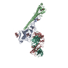
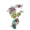
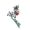
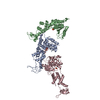
- PDB-1n7u: THE RECEPTOR-BINDING PROTEIN P2 OF BACTERIOPHAGE PRD1: CRYSTAL FORM I -
+ Open data

- Basic information
| Entry | Database: PDB / ID: 1n7u | ||||||
|---|---|---|---|---|---|---|---|
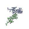
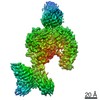
| Title | THE RECEPTOR-BINDING PROTEIN P2 OF BACTERIOPHAGE PRD1: CRYSTAL FORM I | ||||||
 Components Components | Adsorption protein P2 | ||||||
 Keywords Keywords | VIRAL PROTEIN / bacteriophage PRD1 / viral receptor-binding / beta-propeller / proline-rich / antibiotic-resistance | ||||||
| Function / homology |  Function and homology information Function and homology informationvirion component / symbiont entry into host cell / virion attachment to host cell Similarity search - Function | ||||||
| Biological species |   Enterobacteria phage PRD1 (virus) Enterobacteria phage PRD1 (virus) | ||||||
| Method |  X-RAY DIFFRACTION / MAD / Resolution: 2.4 Å X-RAY DIFFRACTION / MAD / Resolution: 2.4 Å | ||||||
 Authors Authors | Xu, L. / Benson, S.D. / Butcher, S.J. / Bamford, D.H. / Burnett, R.M. | ||||||
 Citation Citation | Journal: Structure / Year: 2003 Title: The Receptor Binding Protein P2 of PRD1, a Virus Targeting Antibiotic-Resistant Bacteria, Has a Novel Fold Suggesting Multiple Functions. Authors: Xu, L. / Benson, S.D. / Butcher, S.J. / Bamford, D.H. / Burnett, R.M. #1: Journal: J.STRUCT.BIOL. / Year: 2000Title: Crystallization and preliminary X-ray analysis of receptor-binding protein P2 of bacteriopahge PRD1 Authors: Xu, L. / Butcher, S.J. / Benson, S.D. / Bamford, D.H. / Burnett, R.M. #2: Journal: J.BACTERIOL. / Year: 1999Title: Stable packing of phage PRD1 DNA requires adsorption protein P2, which binds to the IncP plasmid-encoded conjugative transfer complex Authors: Grahn, A.M. / Caldentey, J. / Bamford, J.K.H. / Bamford, D.H. #3: Journal: Adv.Virus Res. / Year: 1995Title: Bacteriophage PRD1: a broad host range dsDNA tectivirus with an internal membrane Authors: Bamford, D.H. / Caldentey, J. / Bamford, J.K.H. #4: Journal: Cell(Cambridge,Mass.) / Year: 1999Title: Viral evolution revealed by bacteriophage PRD1 and human adenovirus coat protein structures Authors: Benson, S.D. / Bamford, J.K.H. / Bamford, D.H. / Burnett, R.M. #5: Journal: J.Mol.Biol. / Year: 1999Title: Bacteriophage PRD1 contains a labile receptor-binding structure at each vertex Authors: Rydman, P.S. / Caldentey, J. / Butcher, S.J. / Fuller, S.D. / Rutten, T. / Bamford, D.H. #6: Journal: Thesis / Year: 2002Title: A tale of two viruses with therapeutic potential: Structural studies on CELO, an avian adenovirus and the bacteriophage PRD1 Authors: Xu, L. | ||||||
| History |
| ||||||
| Remark 351 | The biomolecule is most probably a monomer. Nevertheless, it is notable that the same dimer ... The biomolecule is most probably a monomer. Nevertheless, it is notable that the same dimer appears in the two crystal forms (1N7U and 1N7V). Although the buried surface per subunit in the dimer is ~1,206 A**2, which is relatively substantial, it only represents ~5.5% of the surface area of the P2 monomer. Rate zonal centrifugation, gel filtration, cross-linking, sedimentation equilibrium and low angle X-ray scattering strongly suggest that P2 is a monomer in solution. Furthermore, dynamic light scattering measurements indicated a monodisperse solution with an apparent molecular weight of 79 kDa, which is in reasonable agreement with that for the monomer (63.9 kDa). Nevertheless, P2 may form a loosely associated dimer when bound to the virion. |
- Structure visualization
| Structure viewer | Molecule: MolmilJmol/JSmol |
|---|
- Downloads & links
-Download
| PDBx/mmCIF format | 1n7u.cif.gz | 125 KB | Display | PDBx/mmCIF format |
|---|---|---|---|---|
| PDB format | pdb1n7u.ent.gz | 94.1 KB | Display | PDB format |
| PDBx/mmJSON format | 1n7u.json.gz | Tree view | PDBx/mmJSON format | |
| Others |  Other downloads Other downloads |
-Validation report
| Arichive directory | https://data.pdbj.org/pub/pdb/validation_reports/n7/1n7uftp://data.pdbj.org/pub/pdb/validation_reports/n7/1n7u | HTTPS FTP |
|---|
-Related structure data


-Links
 PDBj
PDBj- Assembly
| Deposited unit | 
| ||||||||
|---|---|---|---|---|---|---|---|---|---|
| 1 | 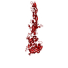
| ||||||||
| Unit cell |
| ||||||||
| Details | Biochemical evidence (gel filtration, low angle X-ray scattering, rate-zonal ultracentrifugation, and dynamic light scattering) indicate that P2 is monomeric, but a potential dimer appears in 3 crystal with different space groups and is obtained for this crystal form by the following symmetry operation: x, -y, -z |
-Components
| #1: Protein | Mass: 59828.297 Da / Num. of mol.: 1 Source method: isolated from a genetically manipulated source Source: (gene. exp.) Enterobacteria phage PRD1 (virus) / Genus: Tectivirus / Gene: II / Plasmid: pMG59 / Production host:  | ||||||
|---|---|---|---|---|---|---|---|
| #2: Chemical |   Mass: 59.044 Da / Num. of mol.: 3 / Source method: obtained synthetically / Formula: C2H3O2 Mass: 59.044 Da / Num. of mol.: 3 / Source method: obtained synthetically / Formula: C2H3O2#3: Chemical | ChemComp-CA / |   Mass: 40.078 Da / Num. of mol.: 1 / Source method: obtained synthetically / Formula: Ca Mass: 40.078 Da / Num. of mol.: 1 / Source method: obtained synthetically / Formula: Ca#4: Water | ChemComp-HOH / |  Mass: 18.015 Da / Num. of mol.: 229 / Source method: isolated from a natural source / Formula: H2O Mass: 18.015 Da / Num. of mol.: 229 / Source method: isolated from a natural source / Formula: H2OHas protein modification | Y | |
-Experimental details
-Experiment
| Experiment | Method: X-RAY DIFFRACTION / Number of used crystals: 1 |
|---|
- Sample preparation
| Crystal | Density Matthews: 3.57 Å3/Da / Density % sol: 65.3 % | ||||||||||||||||||||||||||||
|---|---|---|---|---|---|---|---|---|---|---|---|---|---|---|---|---|---|---|---|---|---|---|---|---|---|---|---|---|---|
| Crystal grow | Temperature: 277 K / Method: vapor diffusion, hanging drop, macroseeding / pH: 4.6 Details: 30% MPD, 0.1 M sodium acetate, 20 mM CaCl2, pH 4.6, VAPOR DIFFUSION, HANGING DROP, MACROSEEDING, temperature 277K | ||||||||||||||||||||||||||||
| Crystal grow | *PLUS Temperature: 4 ℃ / Method: vapor diffusion, hanging drop / Details: used macroseeding | ||||||||||||||||||||||||||||
| Components of the solutions | *PLUS
|
-Data collection
| Diffraction | Mean temperature: 100 K |
|---|---|
| Diffraction source | Source: ROTATING ANODE / Type: RIGAKU RU200 / Wavelength: 1.5418 Å |
| Detector | Type: RIGAKU RAXIS IV / Detector: IMAGE PLATE / Date: Dec 13, 2001 / Details: mirrors |
| Radiation | Monochromator: Yale mirrors / Protocol: SINGLE WAVELENGTH / Monochromatic (M) / Laue (L): M / Scattering type: x-ray |
| Radiation wavelength | Wavelength: 1.5418 Å / Relative weight: 1 |
| Reflection | Resolution: 2.4→43 Å / Num. all: 35306 / Num. obs: 32726 / % possible obs: 92.7 % / Observed criterion σ(F): 1 / Observed criterion σ(I): 1 / Redundancy: 9 % / Biso Wilson estimate: 26.2 Å2 / Rsym value: 0.071 / Net I/σ(I): 12 |
| Reflection shell | Resolution: 2.4→2.55 Å / Num. unique all: 3550 / Rsym value: 0.477 / % possible all: 61.5 |
| Reflection | *PLUS Rmerge(I) obs: 0.071 |
| Reflection shell | *PLUS % possible obs: 78.7 % / Rmerge(I) obs: 0.477 / Mean I/σ(I) obs: 2.1 |
- Processing
| Software |
| ||||||||||||||||||||||||||||||||||||
|---|---|---|---|---|---|---|---|---|---|---|---|---|---|---|---|---|---|---|---|---|---|---|---|---|---|---|---|---|---|---|---|---|---|---|---|---|---|
| Refinement | Method to determine structure: MAD Starting model: Se-Met P2 Resolution: 2.4→43.22 Å / Rfactor Rfree error: 0.007 / Isotropic thermal model: RESTRAINED / Cross valid method: THROUGHOUT / σ(F): 0 / Stereochemistry target values: Engh & Huber
| ||||||||||||||||||||||||||||||||||||
| Solvent computation | Solvent model: FLAT MODEL / Bsol: 32.4238 Å2 / ksol: 0.294665 e/Å3 | ||||||||||||||||||||||||||||||||||||
| Displacement parameters | Biso mean: 46 Å2
| ||||||||||||||||||||||||||||||||||||
| Refine analyze | Luzzati coordinate error free: 0.39 Å / Luzzati sigma a free: 0.44 Å | ||||||||||||||||||||||||||||||||||||
| Refinement step | Cycle: LAST / Resolution: 2.4→43.22 Å
| ||||||||||||||||||||||||||||||||||||
| Refine LS restraints |
| ||||||||||||||||||||||||||||||||||||
| LS refinement shell | Resolution: 2.4→2.55 Å / Rfactor Rfree error: 0.057 / Total num. of bins used: 6
| ||||||||||||||||||||||||||||||||||||
| Xplor file |
| ||||||||||||||||||||||||||||||||||||
| Refinement | *PLUS Highest resolution: 2.4 Å | ||||||||||||||||||||||||||||||||||||
| Solvent computation | *PLUS | ||||||||||||||||||||||||||||||||||||
| Displacement parameters | *PLUS | ||||||||||||||||||||||||||||||||||||
| Refine LS restraints | *PLUS
|
