 Movie
Movie Controller

[English] 日本語
 Yorodumi
- PDB-3n43: Crystal structures of the mature envelope glycoprotein complex (t... -
+ Open data

- Basic information
| Entry | Database: PDB / ID: 3n43 | |||||||||
|---|---|---|---|---|---|---|---|---|---|---|
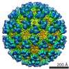
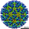
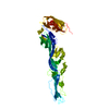
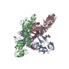
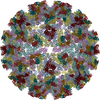
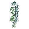
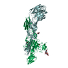
| Title | Crystal structures of the mature envelope glycoprotein complex (trypsin cleavage) of Chikungunya virus. | |||||||||
 Components Components |
| |||||||||
 Keywords Keywords | VIRAL PROTEIN / IMMATURE HETERODIMER / ALPHAVIRUS / RECEPTOR BINDING / MEMBRANE FUSION | |||||||||
| Function / homology |  Function and homology information Function and homology informationT=4 icosahedral viral capsid / host cell endoplasmic reticulum / channel activity / monoatomic ion transmembrane transport / host cell Golgi apparatus / entry receptor-mediated virion attachment to host cell / serine-type endopeptidase activity / fusion of virus membrane with host endosome membrane / symbiont entry into host cell / host cell nucleus ...T=4 icosahedral viral capsid / host cell endoplasmic reticulum / channel activity / monoatomic ion transmembrane transport / host cell Golgi apparatus / entry receptor-mediated virion attachment to host cell / serine-type endopeptidase activity / fusion of virus membrane with host endosome membrane / symbiont entry into host cell / host cell nucleus / host cell plasma membrane / virion membrane / structural molecule activity / proteolysis / RNA binding / identical protein binding Similarity search - Function | |||||||||
| Biological species |   Chikungunya virus Chikungunya virus | |||||||||
| Method |  X-RAY DIFFRACTION / SYNCHROTRON / SAD / Resolution: 2.58 Å X-RAY DIFFRACTION / SYNCHROTRON / SAD / Resolution: 2.58 Å | |||||||||
 Authors Authors | Voss, J. / Vaney, M.C. / Duquerroy, S. / Rey, F.A. | |||||||||
 Citation Citation | Journal: Nature / Year: 2010 Title: Glycoprotein organization of Chikungunya virus particles revealed by X-ray crystallography. Authors: James E Voss / Marie-Christine Vaney / Stéphane Duquerroy / Clemens Vonrhein / Christine Girard-Blanc / Elodie Crublet / Andrew Thompson / Gérard Bricogne / Félix A Rey /  Abstract: Chikungunya virus (CHIKV) is an emerging mosquito-borne alphavirus that has caused widespread outbreaks of debilitating human disease in the past five years. CHIKV invasion of susceptible cells is ...Chikungunya virus (CHIKV) is an emerging mosquito-borne alphavirus that has caused widespread outbreaks of debilitating human disease in the past five years. CHIKV invasion of susceptible cells is mediated by two viral glycoproteins, E1 and E2, which carry the main antigenic determinants and form an icosahedral shell at the virion surface. Glycoprotein E2, derived from furin cleavage of the p62 precursor into E3 and E2, is responsible for receptor binding, and E1 for membrane fusion. In the context of a concerted multidisciplinary effort to understand the biology of CHIKV, here we report the crystal structures of the precursor p62-E1 heterodimer and of the mature E3-E2-E1 glycoprotein complexes. The resulting atomic models allow the synthesis of a wealth of genetic, biochemical, immunological and electron microscopy data accumulated over the years on alphaviruses in general. This combination yields a detailed picture of the functional architecture of the 25 MDa alphavirus surface glycoprotein shell. Together with the accompanying report on the structure of the Sindbis virus E2-E1 heterodimer at acidic pH (ref. 3), this work also provides new insight into the acid-triggered conformational change on the virus particle and its inbuilt inhibition mechanism in the immature complex. #1: Journal: Structure / Year: 2006Title: Structure and interactions at the viral surface of the envelope protein E1 of Semliki Forest virus. Authors: Roussel, A. / Lescar, J. / Vaney, M.C. / Wengler, G. / Rey, F.A. #2: Journal: Nature / Year: 2004Title: Conformational change and protein-protein interactions of the fusion protein of Semliki Forest virus. Authors: Gibbons, D.L. / Vaney, M.C. / Roussel, A. / Vigouroux, A. / Reilly, B. / Lepault, J. / Kielian, M. / Rey, F.A. #3: Journal: Cell(Cambridge,Mass.) / Year: 2001Title: The Fusion glycoprotein shell of Semliki Forest virus: an icosahedral assembly primed for fusogenic activation at endosomal pH. Authors: Lescar, J. / Roussel, A. / Wien, M.W. / Navaza, J. / Fuller, S.D. / Wengler, G. / Rey, F.A. #4: Journal: To be PublishedTitle: Structural Changes of Envelope Proteins During Alphavirus Fusion. Authors: Li, L. / Jose, J. / Xiang, Y. / Kuhn, R.J. / Rossmann, M.G. | |||||||||
| History |
|
- Structure visualization
| Structure viewer | Molecule: MolmilJmol/JSmol |
|---|
- Downloads & links
-Download
| PDBx/mmCIF format | 3n43.cif.gz | 322.7 KB | Display | PDBx/mmCIF format |
|---|---|---|---|---|
| PDB format | pdb3n43.ent.gz | 259.8 KB | Display | PDB format |
| PDBx/mmJSON format | 3n43.json.gz | Tree view | PDBx/mmJSON format | |
| Others |  Other downloads Other downloads |
-Validation report
| Arichive directory | https://data.pdbj.org/pub/pdb/validation_reports/n4/3n43ftp://data.pdbj.org/pub/pdb/validation_reports/n4/3n43 | HTTPS FTP |
|---|
-Related structure data
| Related structure data | 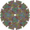 2xfbC 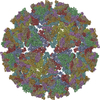 2xfcC  3n40C 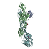 3n41C  3n42C  3n44C C: citing same article ( |
|---|---|
| Similar structure data |


-Links
 PDBj
PDBj


- Assembly
| Deposited unit | 
| ||||||||
|---|---|---|---|---|---|---|---|---|---|
| 1 |
| ||||||||
| Unit cell |
|
-Components
-Protein , 3 types, 3 molecules ABF
| #1: Protein | Mass: 7470.737 Da / Num. of mol.: 1 / Fragment: polyprotein fragment residues 268-318 Source method: isolated from a genetically manipulated source Source: (gene. exp.) Chikungunya virus / Strain: 05-115 / Plasmid: pMRBiP/V5 HisA / Cell line (production host): SCHNEIDER 2 / Production host:  |
|---|---|
| #2: Protein | Mass: 40600.785 Da / Num. of mol.: 1 / Fragment: polyprotein fragment residues 332-667 Source method: isolated from a genetically manipulated source Source: (gene. exp.) Chikungunya virus / Strain: 05-115 / Plasmid: pMRBiP/V5 HisA / Cell line (production host): SCHNEIDER 2 / Production host: |
| #3: Protein | Mass: 50356.223 Da / Num. of mol.: 1 / Fragment: polyprotein fragment residues 810-1200 Source method: isolated from a genetically manipulated source Source: (gene. exp.) Chikungunya virus / Strain: 05-115 / Plasmid: pMRBiP/V5 HisA / Cell line (production host): SCHNEIDER 2 / Production host: |
-Sugars , 2 types, 2 molecules 
| #4: Polysaccharide | 2-acetamido-2-deoxy-alpha-D-glucopyranose-(1-4)-2-acetamido-2-deoxy-beta-D-glucopyranose Source method: isolated from a genetically manipulated source |
|---|---|
| #5: Sugar | ChemComp-NAG /  Type: D-saccharide, beta linking / Mass: 221.208 Da / Num. of mol.: 1 Type: D-saccharide, beta linking / Mass: 221.208 Da / Num. of mol.: 1Source method: isolated from a genetically manipulated source Formula: C8H15NO6 |
-Non-polymers , 2 types, 154 molecules 

| #6: Chemical | ChemComp-ACT /  Mass: 59.044 Da / Num. of mol.: 1 / Source method: obtained synthetically / Formula: C2H3O2 Mass: 59.044 Da / Num. of mol.: 1 / Source method: obtained synthetically / Formula: C2H3O2 |
|---|---|
| #7: Water | ChemComp-HOH / Mass: 18.015 Da / Num. of mol.: 153 / Source method: isolated from a natural source / Formula: H2O |
-Details
| Has protein modification | Y |
|---|---|
| Sequence details | THE RESIDUES ARE PART OF THE LINKER BETWEEN P AND F CHAINS. LINKER WAS CLEAVED BY CHYMOTRYPSIN ...THE RESIDUES ARE PART OF THE LINKER BETWEEN P AND F CHAINS. LINKER WAS CLEAVED BY CHYMOTRYPS |
-Experimental details
-Experiment
| Experiment | Method: X-RAY DIFFRACTION / Number of used crystals: 1 |
|---|
- Sample preparation
| Crystal | Density Matthews: 2.42 Å3/Da / Density % sol: 49.12 % |
|---|---|
| Crystal grow | Temperature: 293 K / pH: 7 Details: 8-12% PEG4K, 100mM NaAcetate, 100mM Hepes pH7.0, VAPOR DIFFUSION, HANGING DROP, temperature 293K |
-Data collection
| Diffraction | Mean temperature: 110 K |
|---|---|
| Diffraction source | Source: SYNCHROTRON / Site: ESRF / Beamline: ID23-2 / Wavelength: 0.8726 |
| Detector | Type: MARMOSAIC 225 mm CCD / Detector: CCD / Date: Sep 19, 2009 Details: ONE PAIR OF (300X40X15) MM3 LONG PT COATED SI MIRROR, 260MM USABLE, IN A KIRKPATRICK-BAEZ GEOMETRY |
| Radiation | Monochromator: SI(111) / Protocol: SINGLE WAVELENGTH / Monochromatic (M) / Laue (L): M / Scattering type: x-ray |
| Radiation wavelength | Wavelength: 0.8726 Å / Relative weight: 1 |
| Reflection | Resolution: 2.58→89.72 Å / % possible obs: 97.7 % / Observed criterion σ(I): 0 / Redundancy: 3.9 % / Biso Wilson estimate: 47.71 Å2 / Rsym value: 0.129 / Net I/σ(I): 8.8 |
| Reflection shell | Resolution: 2.6→2.73 Å / Redundancy: 2 % / Mean I/σ(I) obs: 2 / Rsym value: 0.31 / % possible all: 93.1 |
- Processing
| Software |
| ||||||||||||||||||||||||||||||||||||||||||||||||||||||||||||||||||||||||||||||||||||||||||||||||||||||||||||||||||||||||||||||||||||||||||||||||||||||||||||||||||||||||||||||||||||||||||||||||||||||||||||||||||||||||||||||||||||||||||||||||||||||||||
|---|---|---|---|---|---|---|---|---|---|---|---|---|---|---|---|---|---|---|---|---|---|---|---|---|---|---|---|---|---|---|---|---|---|---|---|---|---|---|---|---|---|---|---|---|---|---|---|---|---|---|---|---|---|---|---|---|---|---|---|---|---|---|---|---|---|---|---|---|---|---|---|---|---|---|---|---|---|---|---|---|---|---|---|---|---|---|---|---|---|---|---|---|---|---|---|---|---|---|---|---|---|---|---|---|---|---|---|---|---|---|---|---|---|---|---|---|---|---|---|---|---|---|---|---|---|---|---|---|---|---|---|---|---|---|---|---|---|---|---|---|---|---|---|---|---|---|---|---|---|---|---|---|---|---|---|---|---|---|---|---|---|---|---|---|---|---|---|---|---|---|---|---|---|---|---|---|---|---|---|---|---|---|---|---|---|---|---|---|---|---|---|---|---|---|---|---|---|---|---|---|---|---|---|---|---|---|---|---|---|---|---|---|---|---|---|---|---|---|---|---|---|---|---|---|---|---|---|---|---|---|---|---|---|---|---|---|---|---|---|---|---|---|---|---|---|---|---|---|---|---|---|
| Refinement | Method to determine structure: SAD Starting model: P62E1 ENVELOPE GLYCOPROTEINS FROM CHIKUNGUNYA VIRUS Resolution: 2.58→44 Å / Cor.coef. Fo:Fc: 0.867 / Cor.coef. Fo:Fc free: 0.862 / SU R Cruickshank DPI: 0.693 / Cross valid method: THROUGHOUT / σ(F): 0 / Stereochemistry target values: Engh & Huber
| ||||||||||||||||||||||||||||||||||||||||||||||||||||||||||||||||||||||||||||||||||||||||||||||||||||||||||||||||||||||||||||||||||||||||||||||||||||||||||||||||||||||||||||||||||||||||||||||||||||||||||||||||||||||||||||||||||||||||||||||||||||||||||
| Displacement parameters | Biso mean: 45.49 Å2
| ||||||||||||||||||||||||||||||||||||||||||||||||||||||||||||||||||||||||||||||||||||||||||||||||||||||||||||||||||||||||||||||||||||||||||||||||||||||||||||||||||||||||||||||||||||||||||||||||||||||||||||||||||||||||||||||||||||||||||||||||||||||||||
| Refine analyze | Luzzati coordinate error obs: 0.48 Å | ||||||||||||||||||||||||||||||||||||||||||||||||||||||||||||||||||||||||||||||||||||||||||||||||||||||||||||||||||||||||||||||||||||||||||||||||||||||||||||||||||||||||||||||||||||||||||||||||||||||||||||||||||||||||||||||||||||||||||||||||||||||||||
| Refinement step | Cycle: LAST / Resolution: 2.58→44 Å
| ||||||||||||||||||||||||||||||||||||||||||||||||||||||||||||||||||||||||||||||||||||||||||||||||||||||||||||||||||||||||||||||||||||||||||||||||||||||||||||||||||||||||||||||||||||||||||||||||||||||||||||||||||||||||||||||||||||||||||||||||||||||||||
| Refine LS restraints |
| ||||||||||||||||||||||||||||||||||||||||||||||||||||||||||||||||||||||||||||||||||||||||||||||||||||||||||||||||||||||||||||||||||||||||||||||||||||||||||||||||||||||||||||||||||||||||||||||||||||||||||||||||||||||||||||||||||||||||||||||||||||||||||
| LS refinement shell | Resolution: 2.58→2.67 Å / Total num. of bins used: 15
| ||||||||||||||||||||||||||||||||||||||||||||||||||||||||||||||||||||||||||||||||||||||||||||||||||||||||||||||||||||||||||||||||||||||||||||||||||||||||||||||||||||||||||||||||||||||||||||||||||||||||||||||||||||||||||||||||||||||||||||||||||||||||||
| Refinement TLS params. | Method: refined / Refine-ID: X-RAY DIFFRACTION
| ||||||||||||||||||||||||||||||||||||||||||||||||||||||||||||||||||||||||||||||||||||||||||||||||||||||||||||||||||||||||||||||||||||||||||||||||||||||||||||||||||||||||||||||||||||||||||||||||||||||||||||||||||||||||||||||||||||||||||||||||||||||||||
| Refinement TLS group |
|
