 Movie
Movie Controller

[English] 日本語
 Yorodumi
- PDB-2xfc: CHIKUNGUNYA E1 E2 ENVELOPE GLYCOPROTEINS FITTED IN SEMLIKI FOREST... -
+ Open data

- Basic information
| Entry | Database: PDB / ID: 2xfc | ||||||
|---|---|---|---|---|---|---|---|
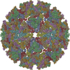
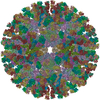
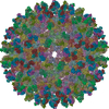
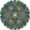
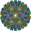
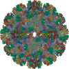
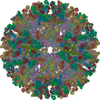
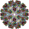
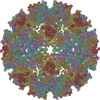
| Title | CHIKUNGUNYA E1 E2 ENVELOPE GLYCOPROTEINS FITTED IN SEMLIKI FOREST VIRUS cryo-EM MAP | ||||||
 Components Components |
| ||||||
 Keywords Keywords | VIRUS / RECEPTOR BINDING / MEMBRANE FUSION / ICOSAHEDRAL ENVELOPED VIRUS | ||||||
| Function / homology |  Function and homology information Function and homology informationT=4 icosahedral viral capsid / host cell endoplasmic reticulum / channel activity / monoatomic ion transmembrane transport / host cell Golgi apparatus / entry receptor-mediated virion attachment to host cell / serine-type endopeptidase activity / fusion of virus membrane with host endosome membrane / symbiont entry into host cell / host cell nucleus ...T=4 icosahedral viral capsid / host cell endoplasmic reticulum / channel activity / monoatomic ion transmembrane transport / host cell Golgi apparatus / entry receptor-mediated virion attachment to host cell / serine-type endopeptidase activity / fusion of virus membrane with host endosome membrane / symbiont entry into host cell / host cell nucleus / host cell plasma membrane / virion membrane / structural molecule activity / proteolysis / RNA binding / identical protein binding Similarity search - Function | ||||||
| Biological species |   CHIKUNGUNYA VIRUS CHIKUNGUNYA VIRUS | ||||||
| Method | ELECTRON MICROSCOPY / single particle reconstruction / cryo EM / Resolution: 9 Å | ||||||
 Authors Authors | Voss, J.E. / Vaney, M.C. / Duquerroy, S. / Rey, F.A. | ||||||
 Citation Citation | Journal: Nature / Year: 2010 Title: Glycoprotein organization of Chikungunya virus particles revealed by X-ray crystallography. Authors: James E Voss / Marie-Christine Vaney / Stéphane Duquerroy / Clemens Vonrhein / Christine Girard-Blanc / Elodie Crublet / Andrew Thompson / Gérard Bricogne / Félix A Rey /  Abstract: Chikungunya virus (CHIKV) is an emerging mosquito-borne alphavirus that has caused widespread outbreaks of debilitating human disease in the past five years. CHIKV invasion of susceptible cells is ...Chikungunya virus (CHIKV) is an emerging mosquito-borne alphavirus that has caused widespread outbreaks of debilitating human disease in the past five years. CHIKV invasion of susceptible cells is mediated by two viral glycoproteins, E1 and E2, which carry the main antigenic determinants and form an icosahedral shell at the virion surface. Glycoprotein E2, derived from furin cleavage of the p62 precursor into E3 and E2, is responsible for receptor binding, and E1 for membrane fusion. In the context of a concerted multidisciplinary effort to understand the biology of CHIKV, here we report the crystal structures of the precursor p62-E1 heterodimer and of the mature E3-E2-E1 glycoprotein complexes. The resulting atomic models allow the synthesis of a wealth of genetic, biochemical, immunological and electron microscopy data accumulated over the years on alphaviruses in general. This combination yields a detailed picture of the functional architecture of the 25 MDa alphavirus surface glycoprotein shell. Together with the accompanying report on the structure of the Sindbis virus E2-E1 heterodimer at acidic pH (ref. 3), this work also provides new insight into the acid-triggered conformational change on the virus particle and its inbuilt inhibition mechanism in the immature complex. #1: Journal: Mol Cell / Year: 2000Title: Cryo-electron microscopy reveals the functional organization of an enveloped virus, Semliki Forest virus. Authors: E J Mancini / M Clarke / B E Gowen / T Rutten / S D Fuller /  Abstract: Semliki Forest virus serves as a paradigm for membrane fusion and assembly. Our icosahedral reconstruction combined 5276 particle images from 48 cryo-electron micrographs and determined the virion ...Semliki Forest virus serves as a paradigm for membrane fusion and assembly. Our icosahedral reconstruction combined 5276 particle images from 48 cryo-electron micrographs and determined the virion structure to 9 A resolution. The improved resolution of this map reveals an N-terminal arm linking capsid subunits and defines the spike-capsid interaction sites. It illustrates the paired helical nature of the transmembrane segments and the elongated structures connecting them to the spike projecting domains. A 10 A diameter density in the fusion protein lines the cavity at the center of the spike. These clearly visible features combine with the variation in order between the layers to provide a framework for understanding the structural changes during the life cycle of an enveloped virus. | ||||||
| History |
|
- Structure visualization
| Movie |
Movie viewer |
|---|---|
| Structure viewer | Molecule: MolmilJmol/JSmol |
 UCSF Chimera
- Downloads & links
-Download
| PDBx/mmCIF format | 2xfc.cif.gz | 571.6 KB | Display | PDBx/mmCIF format |
|---|---|---|---|---|
| PDB format | pdb2xfc.ent.gz | 457.5 KB | Display | PDB format |
| PDBx/mmJSON format | 2xfc.json.gz | Tree view | PDBx/mmJSON format | |
| Others |  Other downloads Other downloads |
-Validation report
| Arichive directory | https://data.pdbj.org/pub/pdb/validation_reports/xf/2xfcftp://data.pdbj.org/pub/pdb/validation_reports/xf/2xfc | HTTPS FTP |
|---|
-Related structure data
| Related structure data | 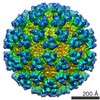 1015M 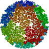 2xfbC 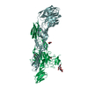 3n40C 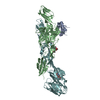 3n41C 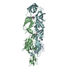 3n42C 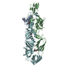 3n43C 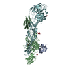 3n44C M: map data used to model this data C: citing same article ( |
|---|---|
| Similar structure data |
-Links
 PDBj
PDBj


- Assembly
| Deposited unit | 
|
|---|---|
| 1 | x 60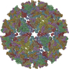
|
| 2 |
|
| 3 | x 5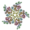
|
| 4 | x 6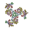
|
| 5 | 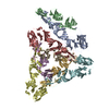
|
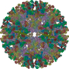
| Symmetry | Point symmetry: (Schoenflies symbol: I (icosahedral)) |
-Components
| #1: Protein | Mass: 47460.934 Da / Num. of mol.: 4 / Fragment: ECTODOMAIN, RESIDUES 810-1248 Source method: isolated from a genetically manipulated source Source: (gene. exp.) CHIKUNGUNYA VIRUS / Strain: 05-115 / Plasmid: PMRBIP/V5HISA / Production host:  #2: Protein | Mass: 47374.086 Da / Num. of mol.: 4 / Fragment: ECTODOMAIN, RESIDUES 326-748 Source method: isolated from a genetically manipulated source Source: (gene. exp.) CHIKUNGUNYA VIRUS / Strain: 05-115 / Plasmid: PMRBIP/V5HISA / Production host: Has protein modification | Y | |
|---|
-Experimental details
-Experiment
| Experiment | Method: ELECTRON MICROSCOPY |
|---|---|
| EM experiment | Aggregation state: PARTICLE / 3D reconstruction method: single particle reconstruction |
- Sample preparation
| Component | Name: semliki forest virus / Type: VIRUS |
|---|---|
| Buffer solution | pH: 7.4 / Details: Tris (10mM) NaCl (100 mM) ph 7.4 |
| Specimen | Embedding applied: NO / Shadowing applied: NO / Staining applied: NO / Vitrification applied: YES |
| Specimen support | Details: OTHER |
| Vitrification | Instrument: HOMEMADE PLUNGER / Cryogen name: ETHANE |
- Electron microscopy imaging
| Microscopy | Model: FEI/PHILIPS CM200FEG/ST / Date: Jan 1, 1995 |
|---|---|
| Electron gun | Electron source: OTHER / Accelerating voltage: 100 kV / Illumination mode: FLOOD BEAM |
| Electron lens | Mode: OTHER / Nominal magnification: 50000 X / Nominal defocus max: 7628 nm / Nominal defocus min: 975 nm |
| Image recording | Electron dose: 8 e/Å2 / Film or detector model: KODAK SO-163 FILM |
| Radiation wavelength | Relative weight: 1 |
- Processing
| Symmetry | Point symmetry: I (icosahedral) | ||||||||||||
|---|---|---|---|---|---|---|---|---|---|---|---|---|---|
| 3D reconstruction | Resolution: 9 Å / Num. of particles: 6000 Details: THE COMPLETE PARTICLE IS GENERATED BY BIOMT MATRICES. THE FIT WAS GENERATED WITH URO IN SINDBIS CRYO-EM MAP EMDB-1015 THAT WAS CORRECTED IN MAGNITUDE BY MULTIPLYING A FACTOR OF 1.038 AND USING PDB ENTRY 3N40 Symmetry type: POINT | ||||||||||||
| Refinement | Highest resolution: 9 Å | ||||||||||||
| Refinement step | Cycle: LAST / Highest resolution: 9 Å
|
