 ムービー
ムービー コントローラー


+ データを開く

- 基本情報
基本情報
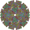
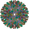
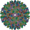
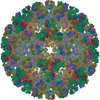
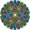
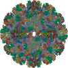
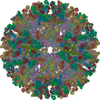
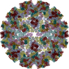
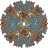
| 登録情報 | データベース: PDB / ID: 2xfc | ||||||
|---|---|---|---|---|---|---|---|
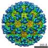
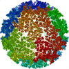
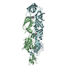
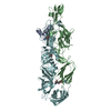
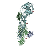
| タイトル | CHIKUNGUNYA E1 E2 ENVELOPE GLYCOPROTEINS FITTED IN SEMLIKI FOREST VIRUS cryo-EM MAP | ||||||
 要素 要素 |
| ||||||
 キーワード キーワード |  VIRUS (ウイルス) / RECEPTOR BINDING (受容体) / MEMBRANE FUSION / ICOSAHEDRAL ENVELOPED VIRUS VIRUS (ウイルス) / RECEPTOR BINDING (受容体) / MEMBRANE FUSION / ICOSAHEDRAL ENVELOPED VIRUS | ||||||
| 機能・相同性 |  機能・相同性情報 機能・相同性情報T=4 icosahedral viral capsid / host cell cytoplasm / symbiont entry into host cell / serine-type endopeptidase activity / fusion of virus membrane with host endosome membrane / structural molecule activity / virion attachment to host cell / host cell plasma membrane / virion membrane / タンパク質分解 ...T=4 icosahedral viral capsid / host cell cytoplasm / symbiont entry into host cell / serine-type endopeptidase activity / fusion of virus membrane with host endosome membrane / structural molecule activity / virion attachment to host cell / host cell plasma membrane / virion membrane / タンパク質分解 / identical protein binding / 細胞膜 / 細胞質類似検索 - 分子機能 | ||||||
| 生物種 |   CHIKUNGUNYA VIRUS (チクングニヤウイルス) CHIKUNGUNYA VIRUS (チクングニヤウイルス) | ||||||
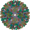
| 手法 | 電子顕微鏡法 / 単粒子再構成法 / クライオ電子顕微鏡法 / 解像度: 9 Å | ||||||
 データ登録者 データ登録者 | Voss, J.E. / Vaney, M.C. / Duquerroy, S. / Rey, F.A. | ||||||
 引用 引用 | ジャーナル: Nature / 年: 2010 タイトル: Glycoprotein organization of Chikungunya virus particles revealed by X-ray crystallography. 著者: James E Voss / Marie-Christine Vaney / Stéphane Duquerroy / Clemens Vonrhein / Christine Girard-Blanc / Elodie Crublet / Andrew Thompson / Gérard Bricogne / Félix A Rey /  要旨: Chikungunya virus (CHIKV) is an emerging mosquito-borne alphavirus that has caused widespread outbreaks of debilitating human disease in the past five years. CHIKV invasion of susceptible cells is ...Chikungunya virus (CHIKV) is an emerging mosquito-borne alphavirus that has caused widespread outbreaks of debilitating human disease in the past five years. CHIKV invasion of susceptible cells is mediated by two viral glycoproteins, E1 and E2, which carry the main antigenic determinants and form an icosahedral shell at the virion surface. Glycoprotein E2, derived from furin cleavage of the p62 precursor into E3 and E2, is responsible for receptor binding, and E1 for membrane fusion. In the context of a concerted multidisciplinary effort to understand the biology of CHIKV, here we report the crystal structures of the precursor p62-E1 heterodimer and of the mature E3-E2-E1 glycoprotein complexes. The resulting atomic models allow the synthesis of a wealth of genetic, biochemical, immunological and electron microscopy data accumulated over the years on alphaviruses in general. This combination yields a detailed picture of the functional architecture of the 25 MDa alphavirus surface glycoprotein shell. Together with the accompanying report on the structure of the Sindbis virus E2-E1 heterodimer at acidic pH (ref. 3), this work also provides new insight into the acid-triggered conformational change on the virus particle and its inbuilt inhibition mechanism in the immature complex. #1: ジャーナル: Mol Cell / 年: 2000タイトル: Cryo-electron microscopy reveals the functional organization of an enveloped virus, Semliki Forest virus. 著者: E J Mancini / M Clarke / B E Gowen / T Rutten / S D Fuller /  要旨: Semliki Forest virus serves as a paradigm for membrane fusion and assembly. Our icosahedral reconstruction combined 5276 particle images from 48 cryo-electron micrographs and determined the virion ...Semliki Forest virus serves as a paradigm for membrane fusion and assembly. Our icosahedral reconstruction combined 5276 particle images from 48 cryo-electron micrographs and determined the virion structure to 9 A resolution. The improved resolution of this map reveals an N-terminal arm linking capsid subunits and defines the spike-capsid interaction sites. It illustrates the paired helical nature of the transmembrane segments and the elongated structures connecting them to the spike projecting domains. A 10 A diameter density in the fusion protein lines the cavity at the center of the spike. These clearly visible features combine with the variation in order between the layers to provide a framework for understanding the structural changes during the life cycle of an enveloped virus. | ||||||
| 履歴 |
|
- 構造の表示
構造の表示
| ムービー |
ムービービューア |
|---|---|
| 構造ビューア | 分子: MolmilJmol/JSmol |
- ダウンロードとリンク
-ダウンロード
| PDBx/mmCIF形式 | 2xfc.cif.gz | 565.5 KB | 表示 | PDBx/mmCIF形式 |
|---|---|---|---|---|
| PDB形式 | pdb2xfc.ent.gz | 466.1 KB | 表示 | PDB形式 |
| PDBx/mmJSON形式 | 2xfc.json.gz | ツリー表示 | PDBx/mmJSON形式 | |
| その他 |  その他のダウンロード その他のダウンロード |
-検証レポート
| アーカイブディレクトリ | https://data.pdbj.org/pub/pdb/validation_reports/xf/2xfcftp://data.pdbj.org/pub/pdb/validation_reports/xf/2xfc | HTTPS FTP |
|---|
-関連構造データ
-リンク
 PDBj
PDBj


- 集合体
集合体
| 登録構造単位 | 
|
|---|---|
| 1 | x 60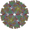
|
| 2 |
|
| 3 | x 5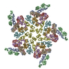
|
| 4 | x 6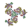
|
| 5 | 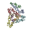
|
| 対称性 | 点対称性: (シェーンフリース記号: I (正20面体型対称)) |
-要素
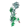
| #1: タンパク質 | 分子量: 47460.934 Da / 分子数: 4 / 断片: ECTODOMAIN, RESIDUES 810-1248 / 由来タイプ: 組換発現 由来: (組換発現) CHIKUNGUNYA VIRUS (チクングニヤウイルス)株: 05-115 / プラスミド: PMRBIP/V5HISA 発現宿主:  DROSOPHILA MELANOGASTER (キイロショウジョウバエ) DROSOPHILA MELANOGASTER (キイロショウジョウバエ)株 (発現宿主): SCHNEIDER 2 / 参照: UniProt: Q1H8W5 #2: タンパク質 | 分子量: 47374.086 Da / 分子数: 4 / 断片: ECTODOMAIN, RESIDUES 326-748 / 由来タイプ: 組換発現 由来: (組換発現) CHIKUNGUNYA VIRUS (チクングニヤウイルス)株: 05-115 / プラスミド: PMRBIP/V5HISA 発現宿主: DROSOPHILA MELANOGASTER (キイロショウジョウバエ)株 (発現宿主): SCHNEIDER 2 / 参照: UniProt: Q1H8W5 |
|---|
-実験情報
-実験
| 実験 | 手法: 電子顕微鏡法 |
|---|---|
| EM実験 | 試料の集合状態: PARTICLE / 3次元再構成法: 単粒子再構成法 |
- 試料調製
試料調製
| 構成要素 | 名称: semliki forest virus / タイプ: VIRUS |
|---|---|
| 緩衝液 | pH: 7.4 / 詳細: Tris (10mM) NaCl (100 mM) ph 7.4 |
| 試料 | 包埋: NO / シャドウイング: NO / 染色: NO / 凍結: YES |
| 試料支持 | 詳細: OTHER |
| 急速凍結 | 装置: HOMEMADE PLUNGER / 凍結剤: ETHANE |
- 電子顕微鏡撮影
| 顕微鏡 | モデル: FEI/PHILIPS CM200FEG/ST / 日付: 1995年1月1日 |
|---|---|
| 電子銃 | 電子線源: その他 / 加速電圧: 100 kV / 照射モード: FLOOD BEAM |
| 電子レンズ | モード: OTHER / 倍率(公称値): 50000 X / 最大 デフォーカス(公称値): 7628 nm / 最小 デフォーカス(公称値): 975 nm |
| 撮影 | 電子線照射量: 8 e/Å2 / フィルム・検出器のモデル: KODAK SO-163 FILM |
| 放射波長 | 相対比: 1 |
- 解析
解析
| 対称性 | 点対称性: I (正20面体型対称) | ||||||||||||
|---|---|---|---|---|---|---|---|---|---|---|---|---|---|
| 3次元再構成 | 解像度: 9 Å / 粒子像の数: 6000 詳細: THE COMPLETE PARTICLE IS GENERATED BY BIOMT MATRICES. THE FIT WAS GENERATED WITH URO IN SINDBIS CRYO-EM MAP EMDB-1015 THAT WAS CORRECTED IN MAGNITUDE BY MULTIPLYING A FACTOR OF 1.038 AND USING PDB ENTRY 3N40 対称性のタイプ: POINT | ||||||||||||
| 精密化 | 最高解像度: 9 Å | ||||||||||||
| 精密化ステップ | サイクル: LAST / 最高解像度: 9 Å
|
