Loading
PDBj
MenuPDBj@FacebookPDBj@TwitterPDBj@YouTubewwPDB FoundationwwPDB
RCSB PDBPDBeBMRBAdv. SearchSearch help

3GBZ
DownloadVisualize
BU of 3gbz by Molmil
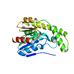
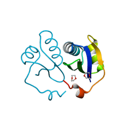
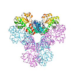
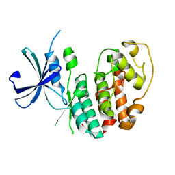
Structure of the CMGC CDK Kinase from Giardia lamblia
Descriptor: Kinase, CMGC CDK
Authors:Seattle Structural Genomics Center for Infectious Disease (SSGCID)
Deposit date:2009-02-20
Release date:2009-03-24
Last modified:2023-09-06
Method:X-RAY DIFFRACTION (1.85 Å)
Cite:Structure of a cyclin-dependent kinase from Giardia lamblia.
Acta Crystallogr.,Sect.F, 67, 2011
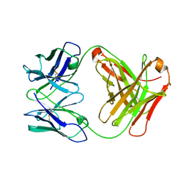
3G5Z
DownloadVisualize
BU of 3g5z by Molmil
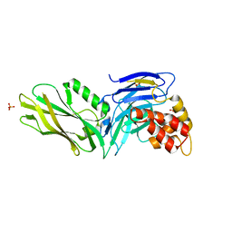
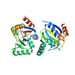
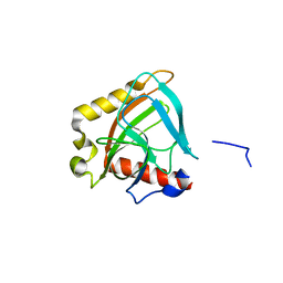
Antibodies Specifically Targeting a Locally Misfolded Region of Tumor Associated EGFR
Descriptor: 175 heavy chain, 175 light chain
Authors:Garrett, T.P.J, Burgess, A.W.
Deposit date:2009-02-05
Release date:2010-02-09
Last modified:2011-10-12
Method:X-RAY DIFFRACTION (2.6 Å)
Cite:Antibodies specifically targeting a locally misfolded region of tumor associated EGFR
Proc.Natl.Acad.Sci.USA, 106, 2009
3G6L
DownloadVisualize
BU of 3g6l by Molmil
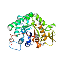
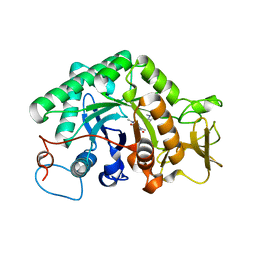
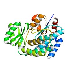
The crystal structure of a chitinase CrChi1 from the nematophagous fungus Clonostachys rosea
Descriptor: Chitinase
Authors:Gan, Z, Lou, Z, Rao, Z, Zhang, K.-Q.
Deposit date:2009-02-06
Release date:2010-02-16
Last modified:2023-11-01
Method:X-RAY DIFFRACTION (1.8 Å)
Cite:Crystal structure and mutagenesis analysis of chitinase CrChi1 from the nematophagous fungus Clonostachys rosea in complex with the inhibitor caffeine
Microbiology, 156, 2010
3DOD
DownloadVisualize
BU of 3dod by Molmil
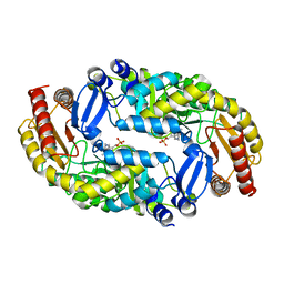
Crystal Structure of PLP Bound 7,8-Diaminopelargonic Acid Synthase in Bacillus subtilis
Descriptor: Adenosylmethionine-8-amino-7-oxononanoate aminotransferase, PYRIDOXAL-5'-PHOSPHATE
Authors:Dey, S, Sacchettini, J.C.
Deposit date:2008-07-03
Release date:2009-06-23
Last modified:2023-08-30
Method:X-RAY DIFFRACTION (1.9 Å)
Cite:Structural characterization of the Mycobacterium tuberculosis biotin biosynthesis enzymes 7,8-diaminopelargonic acid synthase and dethiobiotin synthetase .
Biochemistry, 49, 2010
3DPP
DownloadVisualize
BU of 3dpp by Molmil
Crystal structure of the substrate binding domain of E. coli DnaK in complex with a long pyrrhocoricin-derived inhibitor peptide (form A)
Descriptor: Chaperone protein dnaK, SULFATE ION, inhibitor peptide
Authors:Roujeinikova, A.
Deposit date:2008-07-09
Release date:2009-03-10
Last modified:2023-08-30
Method:X-RAY DIFFRACTION (2.5 Å)
Cite:Allosteric coupling between the lid and interdomain linker in DnaK revealed by inhibitor binding studies.
J.Bacteriol., 191, 2009
3DR6
DownloadVisualize
BU of 3dr6 by Molmil
Structure of yncA, a putative ACETYLTRANSFERASE from Salmonella typhimurium
Descriptor: 1,2-ETHANEDIOL, GLYCEROL, yncA
Authors:Singer, A.U, Skarina, T, Onopriyenko, O, Edwards, A.M, Anderson, W.F, Savchenko, A, Center for Structural Genomics of Infectious Diseases (CSGID)
Deposit date:2008-07-10
Release date:2008-09-09
Last modified:2023-11-15
Method:X-RAY DIFFRACTION (1.75 Å)
Cite:Funded by the national institute of allergy and infectious diseases of nih (contract number hhsn272200700058c).
To be Published
3DYV
DownloadVisualize
BU of 3dyv by Molmil
Snapshots of esterase D from lactobacillus rhamnosus: Insights into a rotation driven catalytic mechanism
Descriptor: CHLORIDE ION, Esterase D
Authors:Bennett, M.D, Delabre, M.-L, Holland, R, Norris, G.E.
Deposit date:2008-07-28
Release date:2009-07-28
Last modified:2023-11-01
Method:X-RAY DIFFRACTION (1.81 Å)
Cite:Snapshots of esterase D from lactobacillus rhamnosus: Insights into a rotation driven catalytic mechanism
To be Published
3GEX
DownloadVisualize
BU of 3gex by Molmil
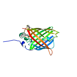
1.6 angstrom crystal structure of fluorescent protein Cypet
Descriptor: Green fluorescent protein
Authors:Hu, X, Liu, R.
Deposit date:2009-02-26
Release date:2010-03-09
Last modified:2023-11-22
Method:X-RAY DIFFRACTION (1.6 Å)
Cite:1.6 angstrom crystal structure of fluorescent protein Cypet
To be Published
3G77
DownloadVisualize
BU of 3g77 by Molmil
Bacterial cytosine deaminase V152A/F316C/D317G mutant
Descriptor: Cytosine deaminase, FE (III) ION
Authors:Stoddard, B, Zhao, L.
Deposit date:2009-02-09
Release date:2009-09-22
Last modified:2024-02-21
Method:X-RAY DIFFRACTION (1.8 Å)
Cite:Bacterial cytosine deaminase mutants created by molecular engineering show improved 5-fluorocytosine-mediated cell killing in vitro and in vivo.
Cancer Res., 69, 2009
3G5B
DownloadVisualize
BU of 3g5b by Molmil
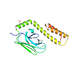
The structure of UNC5b cytoplasmic domain
Descriptor: Netrin receptor UNC5B, PHOSPHATE ION
Authors:Wang, R, Wei, Z, Zhang, M.
Deposit date:2009-02-04
Release date:2009-04-07
Last modified:2024-05-29
Method:X-RAY DIFFRACTION (2 Å)
Cite:Autoinhibition of UNC5b revealed by the cytoplasmic domain structure of the receptor
Mol.Cell, 33, 2009
3G6M
DownloadVisualize
BU of 3g6m by Molmil
crystal structure of a chitinase CrChi1 from the nematophagous fungus Clonostachys rosea in complex with a potent inhibitor caffeine
Descriptor: CAFFEINE, Chitinase
Authors:Gan, Z, Yang, J, Lou, Z, Rao, Z, Zhang, K.-Q.
Deposit date:2009-02-06
Release date:2010-02-16
Last modified:2023-11-01
Method:X-RAY DIFFRACTION (1.65 Å)
Cite:Crystal structure and mutagenesis analysis of chitinase CrChi1 from the nematophagous fungus Clonostachys rosea in complex with the inhibitor caffeine
Microbiology, 156, 2010
3EQS
DownloadVisualize
BU of 3eqs by Molmil
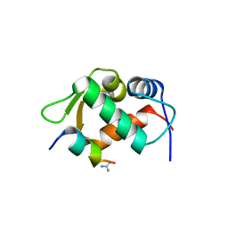
Crystal structure of human MDM2 in complex with a 12-mer peptide inhibitor
Descriptor: 12-mer peptide inhibitor, E3 ubiquitin-protein ligase Mdm2, GUANIDINE
Authors:Pazgier, M, Lu, W.
Deposit date:2008-10-01
Release date:2009-03-17
Last modified:2023-09-06
Method:X-RAY DIFFRACTION (1.65 Å)
Cite:Structural basis for high-affinity peptide inhibition of p53 interactions with MDM2 and MDMX.
Proc.Natl.Acad.Sci.USA, 106, 2009
3ERG
DownloadVisualize
BU of 3erg by Molmil
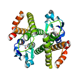
Crystal structure of Gtt2 from Saccharomyces cerevisiae in complex with glutathione sulfnate
Descriptor: GLUTATHIONE SULFONIC ACID, Glutathione S-transferase 2
Authors:Ma, X.X, Jiang, Y.L, He, Y.X, Chen, Y.X, Zhou, C.Z.
Deposit date:2008-10-02
Release date:2009-10-13
Last modified:2023-12-27
Method:X-RAY DIFFRACTION (2.2 Å)
Cite:Structures of yeast glutathione-S-transferase Gtt2 reveal a new catalytic type of GST family.
Embo Rep., 10, 2009
3EUU
DownloadVisualize
BU of 3euu by Molmil
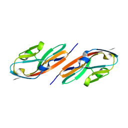
Crystal structure of the FGFR2 D2 domain
Descriptor: Fibroblast growth factor receptor 2
Authors:Sakon, J, Guo, F, Dakshinamurthy, R, Kathir, K.M, Thallapuranam, S.K.K.
Deposit date:2008-10-10
Release date:2009-08-25
Last modified:2023-09-06
Method:X-RAY DIFFRACTION (2.34 Å)
Cite:Crystal structure of the D2 domain of hFGFR2
To be Published
3EWG
DownloadVisualize
BU of 3ewg by Molmil
Crystal structure of the N-terminal domain of NusG (NGN) from Methanocaldococcus jannaschii
Descriptor: 1,4-DIETHYLENE DIOXIDE, Putative transcription antitermination protein nusG
Authors:Zhou, H.H, Liu, Q, Gao, Y.X, Teng, M.K, Niu, L.W.
Deposit date:2008-10-15
Release date:2009-06-16
Last modified:2023-11-01
Method:X-RAY DIFFRACTION (2.04 Å)
Cite:Crystal structure of NusG N-terminal (NGN) domain from Methanocaldococcus jannaschii and its interaction with rpoE''
Proteins, 76, 2009
3EWP
DownloadVisualize
BU of 3ewp by Molmil
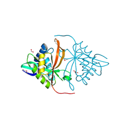
complex of substrate ADP-ribose with IBV Nsp3 ADRP domain
Descriptor: ADENOSINE-5-DIPHOSPHORIBOSE, Non-structural protein 3
Authors:Xu, Y, Cong, L, Chen, C, Wei, L, Zhao, Q, Xu, X, Ma, Y, Bartlam, M, Rao, Z.
Deposit date:2008-10-16
Release date:2009-01-13
Last modified:2023-12-27
Method:X-RAY DIFFRACTION (2 Å)
Cite:Crystal structures of two coronavirus ADP-ribose-1''-monophosphatases and their complexes with ADP-Ribose: a systematic structural analysis of the viral ADRP domain.
J.Virol., 83, 2009
3EZO
DownloadVisualize
BU of 3ezo by Molmil
Crystal structure of acyl-carrier-protein s-malonyltransferase from burkholderia pseudomallei 1710b
Descriptor: Malonyl CoA-acyl carrier protein transacylase
Authors:SSGCID, Seattle Structural Genomics Center for Infectious Disease (SSGCID)
Deposit date:2008-10-23
Release date:2008-11-11
Last modified:2023-09-06
Method:X-RAY DIFFRACTION (2.05 Å)
Cite:Combining functional and structural genomics to sample the essential Burkholderia structome.
Plos One, 8, 2013
3EMK
DownloadVisualize
BU of 3emk by Molmil
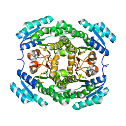
2.5A crystal structure of glucose/ribitol dehydrogenase from brucella melitensis
Descriptor: GLUCOSE/RIBITOL DEHYDROGENASE
Authors:Seattle Structural Genomics Center for Infectious Disease (SSGCID)
Deposit date:2008-09-24
Release date:2008-10-28
Last modified:2024-02-21
Method:X-RAY DIFFRACTION (2.5 Å)
Cite:2.5A crystal structure of glucose/ribitol dehydrogenase from brucella melitensis
To be Published
3EFH
DownloadVisualize
BU of 3efh by Molmil
Crystal structure of human phosphoribosyl pyrophosphate synthetase 1
Descriptor: Ribose-phosphate pyrophosphokinase 1, SULFATE ION
Authors:Li, X, Peng, X, Li, Y.
Deposit date:2008-09-08
Release date:2008-09-30
Last modified:2023-11-01
Method:X-RAY DIFFRACTION (2.6 Å)
Cite:Crystal structure of human phosphoribosyl pyrophosphate synthetase 1
To be Published
3EIZ
DownloadVisualize
BU of 3eiz by Molmil
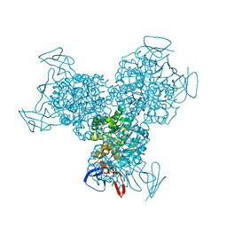
Crystal structure of inorganic pyrophosphatase from burkholderia pseudomallei, H32 crystal form
Descriptor: Inorganic pyrophosphatase
Authors:Seattle Structural Genomics Center for Infectious Disease (SSGCID)
Deposit date:2008-09-17
Release date:2008-09-30
Last modified:2024-02-21
Method:X-RAY DIFFRACTION (1.88 Å)
Cite:Combining functional and structural genomics to sample the essential Burkholderia structome.
Plos One, 8, 2013
3E3E
DownloadVisualize
BU of 3e3e by Molmil
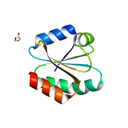
Human Thioredoxin Double Mutant C35S,C73R
Descriptor: HEXAETHYLENE GLYCOL, Thioredoxin
Authors:Hall, G, Emsley, J.
Deposit date:2008-08-07
Release date:2010-03-02
Last modified:2023-08-30
Method:X-RAY DIFFRACTION (2.01 Å)
Cite:Structure of human thioredoxin exhibits a large conformational change.
Protein Sci., 19, 2010
3EOR
DownloadVisualize
BU of 3eor by Molmil
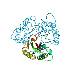
Crystal structure of 2C-methyl-D-erythritol 2,4-clycodiphosphate synthase complexed with ligand
Descriptor: 2-C-methyl-D-erythritol 2,4-cyclodiphosphate synthase, GERANYL DIPHOSPHATE, SODIUM ION, ...
Authors:Hunter, W.N, Ramsden, N.L, Dawson, A.
Deposit date:2008-09-29
Release date:2009-08-25
Last modified:2024-03-20
Method:X-RAY DIFFRACTION (2.9 Å)
Cite:A structure-based approach to ligand discovery for 2C-methyl-D-erythritol-2,4-cyclodiphosphate synthase: a target for antimicrobial therapy
J.Med.Chem., 52, 2009
3EPI
DownloadVisualize
BU of 3epi by Molmil
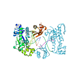
Structure of Human DNA Polymerase Iota complexed with N2-ethylguanine and incoming TTP
Descriptor: 5'-D(*DTP*DCP*DTP*(2EG)P*DGP*DGP*DGP*DTP*DCP*DCP*DTP*DAP*DGP*DGP*DAP*DCP*DCP*(DOC))-3', DNA polymerase iota, PHOSPHATE ION, ...
Authors:Pence, M.G.
Deposit date:2008-09-29
Release date:2008-12-16
Last modified:2023-09-06
Method:X-RAY DIFFRACTION (2.9 Å)
Cite:Lesion bypass of N2-ethylguanine by human DNA polymerase iota.
J.Biol.Chem., 284, 2009
3FA3
DownloadVisualize
BU of 3fa3 by Molmil
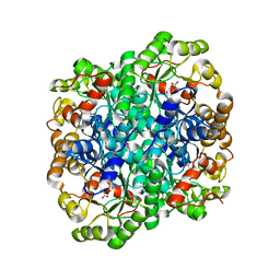
Crystal structure of 2,3-dimethylmalate lyase, a PEP mutase/isocitrate lyase superfamily member, trigonal crystal form
Descriptor: 2,2-difluoro-3,3-dihydroxybutanedioic acid, 2,3-dimethylmalate lyase, GLYCEROL, ...
Authors:Narayanan, B.C, Herzberg, O.
Deposit date:2008-11-14
Release date:2009-01-27
Last modified:2023-12-27
Method:X-RAY DIFFRACTION (2.6 Å)
Cite:Structure and function of 2,3-dimethylmalate lyase, a PEP mutase/isocitrate lyase superfamily member.
J.Mol.Biol., 386, 2009
3FN7
DownloadVisualize
BU of 3fn7 by Molmil
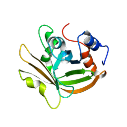
Crystal structure of sortase A (Spy1154) from Streptococcus pyogenes serotype M1 strain SF370
Descriptor: Sortase A
Authors:Banfield, M.J.
Deposit date:2008-12-23
Release date:2009-01-06
Last modified:2023-09-06
Method:X-RAY DIFFRACTION (1.75 Å)
Cite:Crystal Structure of Streptococcus pyogenes Sortase A: Implications for Sortase mechanism
J.Biol.Chem., 284, 2009

223790

數據於2024-08-14公開中

PDB statisticsPDBj update infoContact PDBjnumon